Monday Poster Session
Category: Liver
P2449 - Incidental Neuroendocrine Tumors on Liver Explants
Monday, October 23, 2023
10:30 AM - 4:15 PM PT
Location: Exhibit Hall

Has Audio
- DR
Divya Rayapati, MD
Johns Hopkins Hospital
Baltimore, Maryland
Presenting Author(s)
Divya Rayapati, MD1, Jessica Lin, MD, MBA1, Jacqueline E. Birkness-Gartman, MD2, Kiyoko Oshima, MD, PhD3, Haneyeh Shahbazian, MD4, Ihab RK. Kamel, MD, PhD1, Alia S.. Dadabhai, MD5, Ruhail Kohli, MBBCh4, Kiara Tulla, MD6, Elizabeth King, MD7, Benjamin Philosophe, MD, PhD6, Andrew M. Cameron, MD2, Shane Ottmann, MD1, Ahmet Ö. Gurakar, MD8
1Johns Hopkins Hospital, Baltimore, MD; 2Johns Hopkins University School of Medicine, Baltimore, MD; 3Johns Hopkins University School of medicine, Baltimore, MD; 4Johns Hopkins University, Baltimore, MD; 5Johns Hopkins University, Washington, DC; 6Johns Hopkins, Baltimore, MD; 7Johns Hopkins University, Ellicott City, MD; 8Johns Hopkins, Lutherville, MD
Introduction: Neuroendocrine tumors (NETs), which account for 0.5% of newly diagnosed malignancies, most commonly metastasize to the liver, irrespective of the primary site. Patients undergoing deceased donor liver transplant (DDLT) evaluation receive extensive liver imaging that may reveal incidental hepatic lesions. These lesions are sometimes misdiagnosed as hepatocellular carcinoma (HCC) rather than NETs. We report two cases of patients who underwent DDLT for non-alcoholic steatohepatitis (NASH) and had hepatic lesions on pre-transplant imaging presumed to be HCC. Both lesions were revealed to be NETs upon histopathologic review of liver explants.
Case Description/Methods: Case 1:
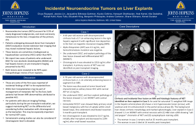
A 65-year-old woman with decompensated cirrhosis had a 0.7 cm enhancing lesion in the right hepatic segment 6 with significant iron deposition in the liver on magnetic resonance imaging (MRI). Alpha fetoprotein (AFP) was 4.5 ng/mL, and hemochromatosis mutation was negative. She underwent DDLT, and explant pathology of the liver lesion showed well-differentiated NET (WDNET). Chromogranin A was elevated to 2253 ng/mL after transplant. A primary source of NET was not identified on post-transplant imaging.
Case 2:
A 70-year-old woman with decompensated cirrhosis had a 1.1 cm arterially enhancing lesion in segment 8 on MRI. The lesion was discussed at tumor board and characterized as solitary lesion HCC with normal AFP at 1.6 ng/mL. She underwent DDLT, and liver explant pathology of the lesion showed 1.2 cm WDNET with central necrosis. Octreotide PET/CT scan showed likely primary small bowel malignancy with foci of uptake within small bowel loops. She underwent small bowel resection 5 months post-transplant. Her chromogranin A was elevated to 5117 ng/mL initially after transplant, and decreased to 2244 ng/mL after small bowel resection.
Both women remain well at 31 and 13 months post-transplant, respectively.
Discussion: These are some of the first cases reported of incidental findings of NET in the explanted liver. While liver transplantation may be part of management of metastatic NET to the liver, both patients had known MRI abnormalities attributed to possible HCC. Thus, for patients with obscure imaging findings, particularly during the pre-transplant evaluation, we suggest maintaining NET on the differential and considering further evaluation with chromogranin A as the biomarker. This may be especially important for normal AFP cases. Somatostatin analog studies can also be considered if chromogranin level is elevated.

Disclosures:
Divya Rayapati, MD1, Jessica Lin, MD, MBA1, Jacqueline E. Birkness-Gartman, MD2, Kiyoko Oshima, MD, PhD3, Haneyeh Shahbazian, MD4, Ihab RK. Kamel, MD, PhD1, Alia S.. Dadabhai, MD5, Ruhail Kohli, MBBCh4, Kiara Tulla, MD6, Elizabeth King, MD7, Benjamin Philosophe, MD, PhD6, Andrew M. Cameron, MD2, Shane Ottmann, MD1, Ahmet Ö. Gurakar, MD8. P2449 - Incidental Neuroendocrine Tumors on Liver Explants, ACG 2023 Annual Scientific Meeting Abstracts. Vancouver, BC, Canada: American College of Gastroenterology.
1Johns Hopkins Hospital, Baltimore, MD; 2Johns Hopkins University School of Medicine, Baltimore, MD; 3Johns Hopkins University School of medicine, Baltimore, MD; 4Johns Hopkins University, Baltimore, MD; 5Johns Hopkins University, Washington, DC; 6Johns Hopkins, Baltimore, MD; 7Johns Hopkins University, Ellicott City, MD; 8Johns Hopkins, Lutherville, MD
Introduction: Neuroendocrine tumors (NETs), which account for 0.5% of newly diagnosed malignancies, most commonly metastasize to the liver, irrespective of the primary site. Patients undergoing deceased donor liver transplant (DDLT) evaluation receive extensive liver imaging that may reveal incidental hepatic lesions. These lesions are sometimes misdiagnosed as hepatocellular carcinoma (HCC) rather than NETs. We report two cases of patients who underwent DDLT for non-alcoholic steatohepatitis (NASH) and had hepatic lesions on pre-transplant imaging presumed to be HCC. Both lesions were revealed to be NETs upon histopathologic review of liver explants.
Case Description/Methods: Case 1:
A 65-year-old woman with decompensated cirrhosis had a 0.7 cm enhancing lesion in the right hepatic segment 6 with significant iron deposition in the liver on magnetic resonance imaging (MRI). Alpha fetoprotein (AFP) was 4.5 ng/mL, and hemochromatosis mutation was negative. She underwent DDLT, and explant pathology of the liver lesion showed well-differentiated NET (WDNET). Chromogranin A was elevated to 2253 ng/mL after transplant. A primary source of NET was not identified on post-transplant imaging.
Case 2:
A 70-year-old woman with decompensated cirrhosis had a 1.1 cm arterially enhancing lesion in segment 8 on MRI. The lesion was discussed at tumor board and characterized as solitary lesion HCC with normal AFP at 1.6 ng/mL. She underwent DDLT, and liver explant pathology of the lesion showed 1.2 cm WDNET with central necrosis. Octreotide PET/CT scan showed likely primary small bowel malignancy with foci of uptake within small bowel loops. She underwent small bowel resection 5 months post-transplant. Her chromogranin A was elevated to 5117 ng/mL initially after transplant, and decreased to 2244 ng/mL after small bowel resection.
Both women remain well at 31 and 13 months post-transplant, respectively.
Discussion: These are some of the first cases reported of incidental findings of NET in the explanted liver. While liver transplantation may be part of management of metastatic NET to the liver, both patients had known MRI abnormalities attributed to possible HCC. Thus, for patients with obscure imaging findings, particularly during the pre-transplant evaluation, we suggest maintaining NET on the differential and considering further evaluation with chromogranin A as the biomarker. This may be especially important for normal AFP cases. Somatostatin analog studies can also be considered if chromogranin level is elevated.

Figure: Cirrhosis and incidental liver lesion on MRI and histologic features of NET identified on liver explant in Case 2: An axial fat-saturated T1-weighted MR image in the hepatic arterial phase (A) shows a 1cm hypervascular lesion (arrow), with washout and rim enhancement in the portal venous phase (B). The background liver shows cirrhotic morphology and marked splenomegaly. Liver explant in Case 2 showed 1.2 cm WDNET with central necrosis with (C) H&E stain x200 showing "salt and pepper" chromatin of NET and (D) synaptophysin staining x200.
Disclosures:
Divya Rayapati indicated no relevant financial relationships.
Jessica Lin indicated no relevant financial relationships.
Jacqueline Birkness-Gartman indicated no relevant financial relationships.
Kiyoko Oshima indicated no relevant financial relationships.
Haneyeh Shahbazian indicated no relevant financial relationships.
Ihab Kamel indicated no relevant financial relationships.
Alia Dadabhai indicated no relevant financial relationships.
Ruhail Kohli indicated no relevant financial relationships.
Kiara Tulla indicated no relevant financial relationships.
Elizabeth King indicated no relevant financial relationships.
Benjamin Philosophe indicated no relevant financial relationships.
Andrew Cameron indicated no relevant financial relationships.
Shane Ottmann indicated no relevant financial relationships.
Ahmet Gurakar indicated no relevant financial relationships.
Divya Rayapati, MD1, Jessica Lin, MD, MBA1, Jacqueline E. Birkness-Gartman, MD2, Kiyoko Oshima, MD, PhD3, Haneyeh Shahbazian, MD4, Ihab RK. Kamel, MD, PhD1, Alia S.. Dadabhai, MD5, Ruhail Kohli, MBBCh4, Kiara Tulla, MD6, Elizabeth King, MD7, Benjamin Philosophe, MD, PhD6, Andrew M. Cameron, MD2, Shane Ottmann, MD1, Ahmet Ö. Gurakar, MD8. P2449 - Incidental Neuroendocrine Tumors on Liver Explants, ACG 2023 Annual Scientific Meeting Abstracts. Vancouver, BC, Canada: American College of Gastroenterology.
