Monday Poster Session
Category: Liver
P2462 - Hepatopleural Fistula & Xanthogranulomatous Infiltrative Liver Mass, Malignancy or Abscess?
Monday, October 23, 2023
10:30 AM - 4:15 PM PT
Location: Exhibit Hall

Has Audio

Samantha Rubin, DO
Lenox Hill Hospital, Northwell Health
New York, NY
Presenting Author(s)
Samantha Rubin, DO, Sunder Sham, MBBS, Noah Mahpour, MD, Hatef Massoumi, MD
Lenox Hill Hospital, Northwell Health, New York, NY
Introduction: Hepatopulmonary fistulas are an uncommon condition characterized by an abnormal connection between the liver and lung. This rarity can arise from various etiologies including infection, trauma, biliary tract obstruction, or surgical complication. Cases of hepatopulmonary fistula stemming from a primary liver etiology are scarce in existing literature. We present a unique case of fistula formation in the right lung pleura caused by an atypical liver abscess, notable for its unusually benign patient presentation.
Case Description/Methods: A 66-year-old female with a history of treated HCV presented to the hospital with three weeks of right upper quadrant discomfort and weight loss. On arrival, she was afebrile with stable vital signs. Laboratory studies revealed WBC 8.79 mm3, Albumin 2.9 g/dL, ALP 163 U/L, AST 17 U/L, ALT 14 U/L, and Bilirubin 0.3 mg/dL. Imaging revealed a 7.7x5.2cm heterogeneously enhancing mass at the subdiaphragmatic posterior right liver and a 5x2.5cm cavitary lung lesion at the posterior right lower lobe with fistulous communication to the liver (A). With concern for hepatic abscess, the patient was started on antibiotics. Infectious work-up for HIV, TB, Entamoeba histolytica, and Echinococcus were negative. Tumor markers of CA 19-9, CEA, and AFP were normal. MRCP showed a right hepatic indeterminate mass without typical features of liver abscess. After discussion with the multidisciplinary tumor board, a liver biopsy was performed to rule out malignancy, which revealed xanthogranulomatous inflammation (XGI) without evidence of carcinoma (B). Tissue culture had no growth of bacteria nor fungus. A CT scan one month later showed slight improvement (C). The patient was subsequently discharged on a prolonged course of antibiotics, with interval imaging planned for ongoing evaluation.
Discussion: A hepatopulmonary fistula poses a considerable mortality risk, reaching up to 10%. Existing literature portrays cases with fever, abdominal pain, and bile-stained sputum. In this case, the patient presented without significant infectious signs. This atypical presentation underscores the importance of conducting a comprehensive diagnostic workup. XGI is an uncommon variant of chronic inflammation that manifests as a destructive tumor-like mass. This is noteworthy as no other similar cases have been reported in the literature. Given the scarcity of information surrounding the occurrence and treatment of this condition, we present this case to contribute to future research and understanding.

Disclosures:
Samantha Rubin, DO, Sunder Sham, MBBS, Noah Mahpour, MD, Hatef Massoumi, MD. P2462 - Hepatopleural Fistula & Xanthogranulomatous Infiltrative Liver Mass, Malignancy or Abscess?, ACG 2023 Annual Scientific Meeting Abstracts. Vancouver, BC, Canada: American College of Gastroenterology.
Lenox Hill Hospital, Northwell Health, New York, NY
Introduction: Hepatopulmonary fistulas are an uncommon condition characterized by an abnormal connection between the liver and lung. This rarity can arise from various etiologies including infection, trauma, biliary tract obstruction, or surgical complication. Cases of hepatopulmonary fistula stemming from a primary liver etiology are scarce in existing literature. We present a unique case of fistula formation in the right lung pleura caused by an atypical liver abscess, notable for its unusually benign patient presentation.
Case Description/Methods: A 66-year-old female with a history of treated HCV presented to the hospital with three weeks of right upper quadrant discomfort and weight loss. On arrival, she was afebrile with stable vital signs. Laboratory studies revealed WBC 8.79 mm3, Albumin 2.9 g/dL, ALP 163 U/L, AST 17 U/L, ALT 14 U/L, and Bilirubin 0.3 mg/dL. Imaging revealed a 7.7x5.2cm heterogeneously enhancing mass at the subdiaphragmatic posterior right liver and a 5x2.5cm cavitary lung lesion at the posterior right lower lobe with fistulous communication to the liver (A). With concern for hepatic abscess, the patient was started on antibiotics. Infectious work-up for HIV, TB, Entamoeba histolytica, and Echinococcus were negative. Tumor markers of CA 19-9, CEA, and AFP were normal. MRCP showed a right hepatic indeterminate mass without typical features of liver abscess. After discussion with the multidisciplinary tumor board, a liver biopsy was performed to rule out malignancy, which revealed xanthogranulomatous inflammation (XGI) without evidence of carcinoma (B). Tissue culture had no growth of bacteria nor fungus. A CT scan one month later showed slight improvement (C). The patient was subsequently discharged on a prolonged course of antibiotics, with interval imaging planned for ongoing evaluation.
Discussion: A hepatopulmonary fistula poses a considerable mortality risk, reaching up to 10%. Existing literature portrays cases with fever, abdominal pain, and bile-stained sputum. In this case, the patient presented without significant infectious signs. This atypical presentation underscores the importance of conducting a comprehensive diagnostic workup. XGI is an uncommon variant of chronic inflammation that manifests as a destructive tumor-like mass. This is noteworthy as no other similar cases have been reported in the literature. Given the scarcity of information surrounding the occurrence and treatment of this condition, we present this case to contribute to future research and understanding.

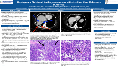
Figure: A: CT of the Abdomen and Pelvis showing a 7.6 x 5.2 x 7.7 cm heterogeneously enhancing mass in hepatic segments 6 and 7 with a thin enhancing rim (blue arrows). A small pocket of gas posterior to this mass tracks superiorly into the cavitary lesion at the posterior right lung base. Central hypo-enhancement within the mass is suggestive of necrosis. A 2.5 x 5.0 cm cavitary lesion in the right lower lobe (red arrows). Gas within the cavitary lesion extends through the right diaphragm to involve the liver.
B: Liver core biopsy showing foamy macrophages/histiocytes arranged in nests, trabeculae, and thickened cords. Chronic inflammatory cells (lymphocytes and plasma cells), fibrosis admixed with foamy histiocytes. There are a few clusters of hepatocytes and bile ducts entrapped within infiltrates of foamy histiocytes. (H&E, 20X)
C: CT of the Abdomen and Pelvis one month after initial imaging, demonstrating a slightly decreased liver lesion and slightly decreased adjacent cavitary lung abscess in posterior right lower lobe.
Disclosures:
Samantha Rubin indicated no relevant financial relationships.
Sunder Sham indicated no relevant financial relationships.
Noah Mahpour indicated no relevant financial relationships.
Hatef Massoumi indicated no relevant financial relationships.
Samantha Rubin, DO, Sunder Sham, MBBS, Noah Mahpour, MD, Hatef Massoumi, MD. P2462 - Hepatopleural Fistula & Xanthogranulomatous Infiltrative Liver Mass, Malignancy or Abscess?, ACG 2023 Annual Scientific Meeting Abstracts. Vancouver, BC, Canada: American College of Gastroenterology.
