Monday Poster Session
Category: Liver
P2469 - Recurrent Hepatocellular Carcinoma Post-Liver Transplantation With Left Atrial Cardiac Metastases
Monday, October 23, 2023
10:30 AM - 4:15 PM PT
Location: Exhibit Hall


Hamdan AlAwadhi, MD
Cleveland Clinic Abu Dhabi
Abu Dhabi, Abu Dhabi, United Arab Emirates
Presenting Author(s)
Hamdan AlAwadhi, MD1, Shiva Kumar, MD2
1Cleveland Clinic Abu Dhabi, Abu Dhabi, Abu Dhabi, United Arab Emirates; 2Digestive Disease Institute, Cleveland Clinic Abu Dhabi, Abu Dhabi, Abu Dhabi, United Arab Emirates
Introduction: Isolated cardiac metastasis of Hepatocellular carcinoma (HCC) discontinuous from the primary liver lesion is extremely rare. We describe the first reported case of post-transplant recurrent HCC presenting with left atrial cardiac metastasis.
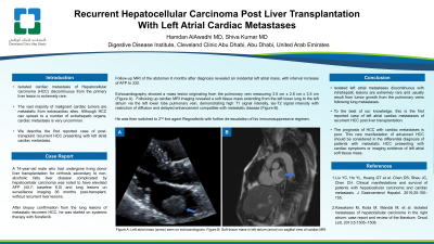
Case Description/Methods: A 74-year-old male who had undergone living donor liver transplantation for cirrhosis secondary to non-alcoholic fatty liver disease complicated by hepatocellular carcinoma was noted to have elevated AFP (44.7; baseline 6.0) and lung lesions on surveillance imaging 36 months post-transplant, without recurrent liver lesions. After biopsy confirmation from the lung lesions of metastatic recurrent HCC, he was started on systemic therapy with Sorafenib. Follow-up MRI of the abdomen 6 months after diagnosis revealed an incidental left atrial mass, with interval increase of AFP to 332. Echocardiography showed a mass lesion originating from the pulmonary vein measuring 3.5 cm x 2.6 cm x 3.5 cm (Figure). Following up cardiac MRI imaging revealed a soft tissue mass extending from the left lower lung to the left atrium via the left lower lobe pulmonary vein, demonstrating high T1 signal intensity, iso-T2 signal intensity with restriction of diffusion and delayed enhancement compatible with metastatic disease (Figure). He was then switched to 2nd line agent Regorafenib with further de-escalation of his immunosuppressive regimen.
Discussion: The vast majority of malignant cardiac tumors are metastatic from extracardiac sites. Although HCC can spread to a number of extrahepatic organs, cardiac metastasis is very uncommon. The majority of reported cardiac metastases of HCC were direct and contiguous extensions of the intrahepatic HCC via the inferior vena cava into the right atrium. Isolated left atrial metastases discontinuous with intrahepatic lesions are extremely rare and usually result from tumor growth from the pulmonary veins following lung metastases. The spectrum of reported clinical manifestations in patients with cardiac metastases of HCC are diverse, ranging from asymptomatic status to chest pain, dyspnea, syncope and heart failure. To the best of our knowledge, this is the first reported case of left atrial cardiac metastases of recurrent HCC post liver transplantation. The prognosis of HCC with cardiac metastases is poor. This rare manifestation of advanced HCC should be considered in the differential diagnosis of patients with metastatic HCC presenting with cardiac symptoms or imaging evidence of left atrial soft tissue mass.

Disclosures:
Hamdan AlAwadhi, MD1, Shiva Kumar, MD2. P2469 - Recurrent Hepatocellular Carcinoma Post-Liver Transplantation With Left Atrial Cardiac Metastases, ACG 2023 Annual Scientific Meeting Abstracts. Vancouver, BC, Canada: American College of Gastroenterology.
1Cleveland Clinic Abu Dhabi, Abu Dhabi, Abu Dhabi, United Arab Emirates; 2Digestive Disease Institute, Cleveland Clinic Abu Dhabi, Abu Dhabi, Abu Dhabi, United Arab Emirates
Introduction: Isolated cardiac metastasis of Hepatocellular carcinoma (HCC) discontinuous from the primary liver lesion is extremely rare. We describe the first reported case of post-transplant recurrent HCC presenting with left atrial cardiac metastasis.
Case Description/Methods: A 74-year-old male who had undergone living donor liver transplantation for cirrhosis secondary to non-alcoholic fatty liver disease complicated by hepatocellular carcinoma was noted to have elevated AFP (44.7; baseline 6.0) and lung lesions on surveillance imaging 36 months post-transplant, without recurrent liver lesions. After biopsy confirmation from the lung lesions of metastatic recurrent HCC, he was started on systemic therapy with Sorafenib. Follow-up MRI of the abdomen 6 months after diagnosis revealed an incidental left atrial mass, with interval increase of AFP to 332. Echocardiography showed a mass lesion originating from the pulmonary vein measuring 3.5 cm x 2.6 cm x 3.5 cm (Figure). Following up cardiac MRI imaging revealed a soft tissue mass extending from the left lower lung to the left atrium via the left lower lobe pulmonary vein, demonstrating high T1 signal intensity, iso-T2 signal intensity with restriction of diffusion and delayed enhancement compatible with metastatic disease (Figure). He was then switched to 2nd line agent Regorafenib with further de-escalation of his immunosuppressive regimen.
Discussion: The vast majority of malignant cardiac tumors are metastatic from extracardiac sites. Although HCC can spread to a number of extrahepatic organs, cardiac metastasis is very uncommon. The majority of reported cardiac metastases of HCC were direct and contiguous extensions of the intrahepatic HCC via the inferior vena cava into the right atrium. Isolated left atrial metastases discontinuous with intrahepatic lesions are extremely rare and usually result from tumor growth from the pulmonary veins following lung metastases. The spectrum of reported clinical manifestations in patients with cardiac metastases of HCC are diverse, ranging from asymptomatic status to chest pain, dyspnea, syncope and heart failure. To the best of our knowledge, this is the first reported case of left atrial cardiac metastases of recurrent HCC post liver transplantation. The prognosis of HCC with cardiac metastases is poor. This rare manifestation of advanced HCC should be considered in the differential diagnosis of patients with metastatic HCC presenting with cardiac symptoms or imaging evidence of left atrial soft tissue mass.

Figure: Echocardiography (left) and Cardiac MRI (right) showing left atrial mass lesion extending from the left pulmonary vein
Disclosures:
Hamdan AlAwadhi indicated no relevant financial relationships.
Shiva Kumar indicated no relevant financial relationships.
Hamdan AlAwadhi, MD1, Shiva Kumar, MD2. P2469 - Recurrent Hepatocellular Carcinoma Post-Liver Transplantation With Left Atrial Cardiac Metastases, ACG 2023 Annual Scientific Meeting Abstracts. Vancouver, BC, Canada: American College of Gastroenterology.
