Monday Poster Session
Category: Liver
P2471 - Rapidly Progressive Combined Hepatocellular-Cholangiocarcinoma
Monday, October 23, 2023
10:30 AM - 4:15 PM PT
Location: Exhibit Hall

Has Audio

Natalie Balassiano, MD
Icahn School of Medicine at Mount Sinai / NYC Health and Hospitals
Queens, NY
Presenting Author(s)
Natalie Balassiano, MD1, Omar Asad, MD2, Merjona Saliaj, MD1
1Icahn School of Medicine at Mount Sinai / NYC Health and Hospitals, Queens, NY; 2University of Miami/HCA Florida JFK Medical Center, Atlantis, FL
Introduction: Combined hepatocellular-cholangiocarcinoma (cHCC-CCA) is a rare primary liver tumor composed of both hepatocytes and biliary ductal epithelium. Risk factors and presenting symptoms match those of other biliary malignancies, however, its incidence is reported in less than 5% of primary liver tumors.
Case Description/Methods: A 76 year old female with a history of obesity, hyperlipidemia, and diabetes mellitus, presented with 4 days of non-bloody non-bilious vomiting and constipation. She denied any history of alcohol or tobacco use. Labs were notable for creatinine of 2.65 mg/dL and alkaline phosphatase (ALP) of 107 U/L. CT of the abdomen and pelvis showed a heterogeneous attenuation within the liver adjacent to the gallbladder and increased attenuation within the gallbladder. Her symptoms were attributed to diabetic gastroparesis and improved with metoclopramide.
The following month, she was readmitted with complaints of weakness, generalized pruritus, abdominal pain, jaundice, and weight loss. Her physical exam was significant for scleral icterus, jaundice, spider angioma, and abdominal tenderness. Labs during this admission showed hypoalbuminemia of 2.9 g/dL, hyperbilirubinemia of 17.6 mg/dL with direct bilirubin of 14.8 mg/dL, ALP 528 U/L, ALT 58 U/L and AST at 170 U/L. Viral and autoimmune hepatitis studies were negative. Tumor markers were significant for alpha-fetoprotein of 4,710 ng/mL, CA 19-9 of 405 U/mL and CA-125 of 119 U/mL. CT of the abdomen revealed cirrhosis with attenuation of the hepatic parenchyma noted on the prior scan (Figure A). MRCP was suspicious for a mass (Figure B). Biopsy of the lesion showed poorly differentiated cHCC-CCA with necrosis. Within 1 month of radiological findings and 15 days of deranged hepatic function, the patient subsequently passed.
Discussion: Although cHCC-CCA is a rare malignancy, it is important for clinicians to be aware of this mixed-type tumor that exhibits features of both hepatocellular carcinoma and cholangiocarcinoma. This tumor should be considered in the appropriate clinical setting, even in patients without traditional risk factors. The unique aspect of this case is the patient's rapid deterioration. In retrospect, an earlier diagnosis through an inpatient gastroenterology consultation may have been beneficial. Despite the high mortality rate associated with cHCC-CCA, an early diagnosis would have allowed for earlier treatment, potentially improving the patient's quality of life and aligning with their goals of care.

Disclosures:
Natalie Balassiano, MD1, Omar Asad, MD2, Merjona Saliaj, MD1. P2471 - Rapidly Progressive Combined Hepatocellular-Cholangiocarcinoma, ACG 2023 Annual Scientific Meeting Abstracts. Vancouver, BC, Canada: American College of Gastroenterology.
1Icahn School of Medicine at Mount Sinai / NYC Health and Hospitals, Queens, NY; 2University of Miami/HCA Florida JFK Medical Center, Atlantis, FL
Introduction: Combined hepatocellular-cholangiocarcinoma (cHCC-CCA) is a rare primary liver tumor composed of both hepatocytes and biliary ductal epithelium. Risk factors and presenting symptoms match those of other biliary malignancies, however, its incidence is reported in less than 5% of primary liver tumors.
Case Description/Methods: A 76 year old female with a history of obesity, hyperlipidemia, and diabetes mellitus, presented with 4 days of non-bloody non-bilious vomiting and constipation. She denied any history of alcohol or tobacco use. Labs were notable for creatinine of 2.65 mg/dL and alkaline phosphatase (ALP) of 107 U/L. CT of the abdomen and pelvis showed a heterogeneous attenuation within the liver adjacent to the gallbladder and increased attenuation within the gallbladder. Her symptoms were attributed to diabetic gastroparesis and improved with metoclopramide.
The following month, she was readmitted with complaints of weakness, generalized pruritus, abdominal pain, jaundice, and weight loss. Her physical exam was significant for scleral icterus, jaundice, spider angioma, and abdominal tenderness. Labs during this admission showed hypoalbuminemia of 2.9 g/dL, hyperbilirubinemia of 17.6 mg/dL with direct bilirubin of 14.8 mg/dL, ALP 528 U/L, ALT 58 U/L and AST at 170 U/L. Viral and autoimmune hepatitis studies were negative. Tumor markers were significant for alpha-fetoprotein of 4,710 ng/mL, CA 19-9 of 405 U/mL and CA-125 of 119 U/mL. CT of the abdomen revealed cirrhosis with attenuation of the hepatic parenchyma noted on the prior scan (Figure A). MRCP was suspicious for a mass (Figure B). Biopsy of the lesion showed poorly differentiated cHCC-CCA with necrosis. Within 1 month of radiological findings and 15 days of deranged hepatic function, the patient subsequently passed.
Discussion: Although cHCC-CCA is a rare malignancy, it is important for clinicians to be aware of this mixed-type tumor that exhibits features of both hepatocellular carcinoma and cholangiocarcinoma. This tumor should be considered in the appropriate clinical setting, even in patients without traditional risk factors. The unique aspect of this case is the patient's rapid deterioration. In retrospect, an earlier diagnosis through an inpatient gastroenterology consultation may have been beneficial. Despite the high mortality rate associated with cHCC-CCA, an early diagnosis would have allowed for earlier treatment, potentially improving the patient's quality of life and aligning with their goals of care.

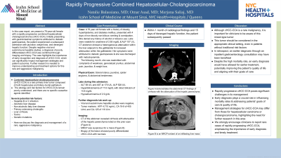
Figure: Image A demonstrates the abdominal CT findings of cirrhosis with the attenuation of the hepatic parenchyma. Image B is an MRCP evident of an infiltrating liver mass.
Disclosures:
Natalie Balassiano indicated no relevant financial relationships.
Omar Asad indicated no relevant financial relationships.
Merjona Saliaj indicated no relevant financial relationships.
Natalie Balassiano, MD1, Omar Asad, MD2, Merjona Saliaj, MD1. P2471 - Rapidly Progressive Combined Hepatocellular-Cholangiocarcinoma, ACG 2023 Annual Scientific Meeting Abstracts. Vancouver, BC, Canada: American College of Gastroenterology.
