Monday Poster Session
Category: Liver
P2477 - β-Catenin Activated Hepatocellular Carcinoma in a Young Man
Monday, October 23, 2023
10:30 AM - 4:15 PM PT
Location: Exhibit Hall

Has Audio
- LX
Lankai C. Xu, MD, MS
University of California Davis Medical Center
Sacramento, CA
Presenting Author(s)
Lankai C. Xu, MD, MS, Vikrant Rachakonda, MD
University of California Davis Medical Center, Sacramento, CA
Introduction: Hepatocellular adenoma (HCA) is a benign and rare liver tumor that most commonly presents in patients with chronic liver disease from hepatitis or cirrhosis. Risk factors for HCA include exposure to oral contraceptives, hormone replacement therapy, and glycogen storage disease. Resection of HCA is recommended because of risk of hemorrhage or malignant transformation. Risk factors for malignancy include male sex, tumor size >5cm, and β-catenin-activated HCA.
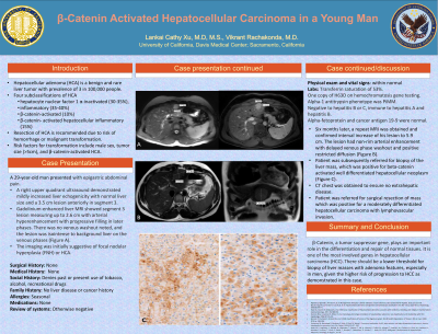
Case Description/Methods: A 29-year-old man was referred for evaluation of epigastric abdominal pain. A right upper quadrant ultrasound demonstrated a 3.5 cm lesion anteriorly in segment 3. He underwent a gadolinium-enhanced liver MRI notable for a segment 3 lesion measuring up to 2.6 cm with arterial hyperenhancement with progressive filling in later phases. There was no venous washout noted, and the lesion was isointense to background liver on the venous phases. The imaging was initially suggestive of focal nodular hyperplasia (FNH) or HCA. The patient had no past medical history or family history of any liver diseases. He denied alcohol, tobacco, illicit substance use, and blood transfusions. Physical examination was within normal. Laboratory data was notable for elevated transferrin saturation of 53%, but with only one copy of H63D on hemochromatosis gene testing. Alpha-1 antitrypsin phenotype was PiMM. He had no evidence of viral hepatitis B or C, and he was immune to hepatitis A and B. Alpha-fetoprotein and cancer antigen 19-9 were normal.
Six months later, a surveillance ultrasound revealed an increase in size of the lesion to 5.8 cm. Repeat MRI confirmed interval increase of the lesion to 5.9 cm. The lesion had non-rim arterial enhancement with delayed venous phase washout and positive restricted diffusion. Liver mass biopsy was positive for beta-catenin activated well differentiated hepatocellular neoplasm. CT chest did not show extrahepatic disease. Patient was referred for surgical resection of mass which was positive for a moderately differentiated hepatocellular carcinoma with lymphovascular invasion.
Discussion: β-Catenin plays an important role in the differentiation and repair of normal tissues. It is one of the most involved genes in hepatocellular carcinoma (HCC). There should be a lower threshold for biopsy of liver masses with adenoma features, especially in men, given the higher risk of progression to HCC.
Disclosures:
Lankai C. Xu, MD, MS, Vikrant Rachakonda, MD. P2477 - β-Catenin Activated Hepatocellular Carcinoma in a Young Man, ACG 2023 Annual Scientific Meeting Abstracts. Vancouver, BC, Canada: American College of Gastroenterology.
University of California Davis Medical Center, Sacramento, CA
Introduction: Hepatocellular adenoma (HCA) is a benign and rare liver tumor that most commonly presents in patients with chronic liver disease from hepatitis or cirrhosis. Risk factors for HCA include exposure to oral contraceptives, hormone replacement therapy, and glycogen storage disease. Resection of HCA is recommended because of risk of hemorrhage or malignant transformation. Risk factors for malignancy include male sex, tumor size >5cm, and β-catenin-activated HCA.
Case Description/Methods: A 29-year-old man was referred for evaluation of epigastric abdominal pain. A right upper quadrant ultrasound demonstrated a 3.5 cm lesion anteriorly in segment 3. He underwent a gadolinium-enhanced liver MRI notable for a segment 3 lesion measuring up to 2.6 cm with arterial hyperenhancement with progressive filling in later phases. There was no venous washout noted, and the lesion was isointense to background liver on the venous phases. The imaging was initially suggestive of focal nodular hyperplasia (FNH) or HCA. The patient had no past medical history or family history of any liver diseases. He denied alcohol, tobacco, illicit substance use, and blood transfusions. Physical examination was within normal. Laboratory data was notable for elevated transferrin saturation of 53%, but with only one copy of H63D on hemochromatosis gene testing. Alpha-1 antitrypsin phenotype was PiMM. He had no evidence of viral hepatitis B or C, and he was immune to hepatitis A and B. Alpha-fetoprotein and cancer antigen 19-9 were normal.
Six months later, a surveillance ultrasound revealed an increase in size of the lesion to 5.8 cm. Repeat MRI confirmed interval increase of the lesion to 5.9 cm. The lesion had non-rim arterial enhancement with delayed venous phase washout and positive restricted diffusion. Liver mass biopsy was positive for beta-catenin activated well differentiated hepatocellular neoplasm. CT chest did not show extrahepatic disease. Patient was referred for surgical resection of mass which was positive for a moderately differentiated hepatocellular carcinoma with lymphovascular invasion.
Discussion: β-Catenin plays an important role in the differentiation and repair of normal tissues. It is one of the most involved genes in hepatocellular carcinoma (HCC). There should be a lower threshold for biopsy of liver masses with adenoma features, especially in men, given the higher risk of progression to HCC.
Disclosures:
Lankai Xu indicated no relevant financial relationships.
Vikrant Rachakonda indicated no relevant financial relationships.
Lankai C. Xu, MD, MS, Vikrant Rachakonda, MD. P2477 - β-Catenin Activated Hepatocellular Carcinoma in a Young Man, ACG 2023 Annual Scientific Meeting Abstracts. Vancouver, BC, Canada: American College of Gastroenterology.
