Monday Poster Session
Category: Liver
P2488 - A Rare Case of Small Cell Carcinoma of Liver
Monday, October 23, 2023
10:30 AM - 4:15 PM PT
Location: Exhibit Hall

Has Audio

Bikal Lamichhane, MBBS
Guthrie Robert Packer Hospital
Sayre, PA
Presenting Author(s)
Bikal Lamichhane, MBBS1, Sudhir Pasham, MBBS2, Sandhya Dhital, MD3, Saral Lamichhane, MBBS4
1Guthrie Robert Packer Hospital, Sayre, PA; 2Robert Packer Hospital, Sayre, PA; 3University of California, Castro Valley, CA; 4Woodhull Hospital, Middle Village, NY
Introduction: Small cell carcinoma (SCC) usually occurs in the lungs, where they account for 25% of lung carcinomas. Extrapulmonary small cell carcinomas (EPSCC) are rare aggressive neoplasms, accounting for only 2-4% of SCCs. Almost half of extrapulmonary cases are found in the gastrointestinal tract. Primary SCC in the hepatobiliary system has been reported in only a few reported cases in the literature.
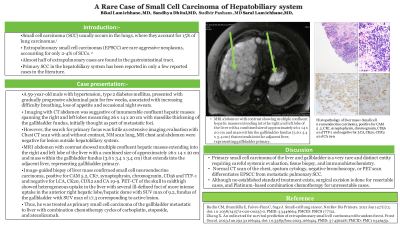
Case Description/Methods: A 59-year-old male with hypertension, type 2 diabetes mellitus, presented with gradually progressive abdominal pain for few weeks, associated with increasing difficulty breathing, loss of appetite and occasional night sweats. Imaging with CT abdomen was suggestive of innumerable confluent hepatic masses spanning the right and left lobes measuring 26 x 14 x 20 cm with masslike thickening of the gallbladder fundus, initially thought as part of metastatic foci. However, the search for primary focus was futile as extensive imaging evaluation with Chest CT scan with and without contrast, NM scan lung, MR chest and abdomen were negative for lesion outside hepatobiliary system. MRI abdomen with contrast showed multiple confluent hepatic masses extending into the right and left lobe of the liver with a combined size of approximately 26 x 14 x 20 cm and mass within the gallbladder fundus (3.6 x 3.4 x 3.4 cm) that extends into the adjacent liver, representing gallbladder primary. Image-guided biopsy of liver mass confirmed small cell neuroendocrine carcinoma, positive for CAM 5.2, CK7, synaptophysin, chromogranin, CD56 and TTF-1 and negative for LCA, CK20, CDX2 and CA 19-9. PET-CT of the skull to midthigh showed heterogeneous uptake in the liver with several ill-defined foci of more intense uptake in the anterior right hepatic lobe/hepatic dome with SUV max of 9.2, fundus of the gallbladder with SUV max of 17.3 corresponding to active lesion. Thus, he was treated as primary small cell carcinoma of the gallbladder metastatic to liver with combination chemotherapy cycles of carboplatin, etoposide, and atezolizumab.
Discussion: Primary small cell carcinoma of the liver and gallbladder is a very rare and distinct entity requiring careful systemic evaluation, tissue biopsy, and immunohistochemistry. Normal CT scan of the chest, sputum cytology, negative bronchoscopy, or PET scan differentiates EPSCC from metastatic pulmonary SCC. Although no established standard treatment exists, surgical excision is done for resectable cases, and Platinum-based combination chemotherapy for unresectable cases.

Disclosures:
Bikal Lamichhane, MBBS1, Sudhir Pasham, MBBS2, Sandhya Dhital, MD3, Saral Lamichhane, MBBS4. P2488 - A Rare Case of Small Cell Carcinoma of Liver, ACG 2023 Annual Scientific Meeting Abstracts. Vancouver, BC, Canada: American College of Gastroenterology.
1Guthrie Robert Packer Hospital, Sayre, PA; 2Robert Packer Hospital, Sayre, PA; 3University of California, Castro Valley, CA; 4Woodhull Hospital, Middle Village, NY
Introduction: Small cell carcinoma (SCC) usually occurs in the lungs, where they account for 25% of lung carcinomas. Extrapulmonary small cell carcinomas (EPSCC) are rare aggressive neoplasms, accounting for only 2-4% of SCCs. Almost half of extrapulmonary cases are found in the gastrointestinal tract. Primary SCC in the hepatobiliary system has been reported in only a few reported cases in the literature.
Case Description/Methods: A 59-year-old male with hypertension, type 2 diabetes mellitus, presented with gradually progressive abdominal pain for few weeks, associated with increasing difficulty breathing, loss of appetite and occasional night sweats. Imaging with CT abdomen was suggestive of innumerable confluent hepatic masses spanning the right and left lobes measuring 26 x 14 x 20 cm with masslike thickening of the gallbladder fundus, initially thought as part of metastatic foci. However, the search for primary focus was futile as extensive imaging evaluation with Chest CT scan with and without contrast, NM scan lung, MR chest and abdomen were negative for lesion outside hepatobiliary system. MRI abdomen with contrast showed multiple confluent hepatic masses extending into the right and left lobe of the liver with a combined size of approximately 26 x 14 x 20 cm and mass within the gallbladder fundus (3.6 x 3.4 x 3.4 cm) that extends into the adjacent liver, representing gallbladder primary. Image-guided biopsy of liver mass confirmed small cell neuroendocrine carcinoma, positive for CAM 5.2, CK7, synaptophysin, chromogranin, CD56 and TTF-1 and negative for LCA, CK20, CDX2 and CA 19-9. PET-CT of the skull to midthigh showed heterogeneous uptake in the liver with several ill-defined foci of more intense uptake in the anterior right hepatic lobe/hepatic dome with SUV max of 9.2, fundus of the gallbladder with SUV max of 17.3 corresponding to active lesion. Thus, he was treated as primary small cell carcinoma of the gallbladder metastatic to liver with combination chemotherapy cycles of carboplatin, etoposide, and atezolizumab.
Discussion: Primary small cell carcinoma of the liver and gallbladder is a very rare and distinct entity requiring careful systemic evaluation, tissue biopsy, and immunohistochemistry. Normal CT scan of the chest, sputum cytology, negative bronchoscopy, or PET scan differentiates EPSCC from metastatic pulmonary SCC. Although no established standard treatment exists, surgical excision is done for resectable cases, and Platinum-based combination chemotherapy for unresectable cases.

Figure: MRI abdomen with contrast showing multiple confluent hepatic masses extending into the right and left lobe of the liver with a combined size of approximately 26 x 14 x 20 cm and mass within the gallbladder fundus (3.6 x 3.4 x 3.4 cm) that extends into the adjacent liver, representing gallbladder primary.
Disclosures:
Bikal Lamichhane indicated no relevant financial relationships.
Sudhir Pasham indicated no relevant financial relationships.
Sandhya Dhital indicated no relevant financial relationships.
Saral Lamichhane indicated no relevant financial relationships.
Bikal Lamichhane, MBBS1, Sudhir Pasham, MBBS2, Sandhya Dhital, MD3, Saral Lamichhane, MBBS4. P2488 - A Rare Case of Small Cell Carcinoma of Liver, ACG 2023 Annual Scientific Meeting Abstracts. Vancouver, BC, Canada: American College of Gastroenterology.
