Monday Poster Session
Category: Liver
P2489 - Conquering Ectopic Variceal Bleeding: A Clinical Vignette
Monday, October 23, 2023
10:30 AM - 4:15 PM PT
Location: Exhibit Hall

Has Audio
- MA
Matthew B. Alias, MD, MS
Medical University of South Carolina
Charleston, SC
Presenting Author(s)
Matthew B. Alias, MD, MS, Thomas E. Werth, MD
Medical University of South Carolina, Charleston, SC
Introduction: Ectopic varices, defined as non-gastroesophageal varices occurring in the presence of intra and extrahepatic portal hypertension, account for up to 5% of all variceal bleeding events. Gastroenterologists are presented with a clinical challenge due to significant variations in diagnosis and management of ectopic varices.
Case Description/Methods: A 55-year-old male with a history of homelessness, diverticulitis with colostomy and reversal, hepatitis C, alcohol cirrhosis with ascites, presented due to encephalopathy. Presenting symptoms were 5 days of abdominal pain, black diarrhea, and heavy NSAID and alcohol use. His vitals were notable for hypotension, tachycardia, and hemoglobin of 5.4 g/dL. Presuming a variceal bleed, blood transfusions, octreotide, proton pump inhibitor and ceftriaxone were administered. Esophagogastroduodenoscopy (EGD) showed mild portal hypertensive gastropathy but failed to identify the bleeding source.
Overnight, the patient had extensive melena and he became hemodynamically unstable, necessitating intubation and initiation of a massive transfusion protocol. Due to a contrast shortage, CT angiography (CTA) was unavailable. Repeat EGD, push enteroscopy, and colonoscopy confirmed significant bleeding, but could not locate the source. Consequently, a CTA and venogram were obtained, revealing extensive superior mesenteric vein (SMV) varices. The patient underwent successful direct coil embolization with interventional radiology.
Discussion: Ectopic varices defy the typical presentation and management of gastroesophageal varices, as they occur in various locations such as the small bowel, large bowel, gall bladder/common bile duct, retroperitoneum, and peristomal areas. The varices are associated with increased bleeding risk, especially at lower portal pressures, and have a higher incidence of rebleeding. Unfortunately, there are no clear guidelines on management due to the heterogenous nature of the disease presentation and a paucity of randomized control trials. Treatment options may include injection sclerotherapy, endoscopic band ligation, surgical ligation, and shunting procedures (transjugular intra-hepatic portosystemic shunt (TIPS) and balloon occluded retrograde transvenous obliteration). Due to concerns about patient follow up after discharge, and his intubated state, direct coil embolization was chosen as the preferred treatment modality. This case highlights the importance of a multidisciplinary approach for optimal management of ectopic variceal bleeding.

Disclosures:
Matthew B. Alias, MD, MS, Thomas E. Werth, MD. P2489 - Conquering Ectopic Variceal Bleeding: A Clinical Vignette, ACG 2023 Annual Scientific Meeting Abstracts. Vancouver, BC, Canada: American College of Gastroenterology.
Medical University of South Carolina, Charleston, SC
Introduction: Ectopic varices, defined as non-gastroesophageal varices occurring in the presence of intra and extrahepatic portal hypertension, account for up to 5% of all variceal bleeding events. Gastroenterologists are presented with a clinical challenge due to significant variations in diagnosis and management of ectopic varices.
Case Description/Methods: A 55-year-old male with a history of homelessness, diverticulitis with colostomy and reversal, hepatitis C, alcohol cirrhosis with ascites, presented due to encephalopathy. Presenting symptoms were 5 days of abdominal pain, black diarrhea, and heavy NSAID and alcohol use. His vitals were notable for hypotension, tachycardia, and hemoglobin of 5.4 g/dL. Presuming a variceal bleed, blood transfusions, octreotide, proton pump inhibitor and ceftriaxone were administered. Esophagogastroduodenoscopy (EGD) showed mild portal hypertensive gastropathy but failed to identify the bleeding source.
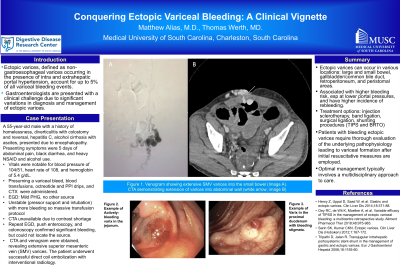
Overnight, the patient had extensive melena and he became hemodynamically unstable, necessitating intubation and initiation of a massive transfusion protocol. Due to a contrast shortage, CT angiography (CTA) was unavailable. Repeat EGD, push enteroscopy, and colonoscopy confirmed significant bleeding, but could not locate the source. Consequently, a CTA and venogram were obtained, revealing extensive superior mesenteric vein (SMV) varices. The patient underwent successful direct coil embolization with interventional radiology.
Discussion: Ectopic varices defy the typical presentation and management of gastroesophageal varices, as they occur in various locations such as the small bowel, large bowel, gall bladder/common bile duct, retroperitoneum, and peristomal areas. The varices are associated with increased bleeding risk, especially at lower portal pressures, and have a higher incidence of rebleeding. Unfortunately, there are no clear guidelines on management due to the heterogenous nature of the disease presentation and a paucity of randomized control trials. Treatment options may include injection sclerotherapy, endoscopic band ligation, surgical ligation, and shunting procedures (transjugular intra-hepatic portosystemic shunt (TIPS) and balloon occluded retrograde transvenous obliteration). Due to concerns about patient follow up after discharge, and his intubated state, direct coil embolization was chosen as the preferred treatment modality. This case highlights the importance of a multidisciplinary approach for optimal management of ectopic variceal bleeding.

Figure: Figures: Venogram showing extensive SMV varices into the small bowel (image A). CTA demonstrating extension of varices into abdominal wall (white arrow; image B)
Disclosures:
Matthew Alias indicated no relevant financial relationships.
Thomas Werth indicated no relevant financial relationships.
Matthew B. Alias, MD, MS, Thomas E. Werth, MD. P2489 - Conquering Ectopic Variceal Bleeding: A Clinical Vignette, ACG 2023 Annual Scientific Meeting Abstracts. Vancouver, BC, Canada: American College of Gastroenterology.
