Monday Poster Session
Category: Liver
P2506 - A Case of Primary Hepatic Sarcoma Presenting as a Hepatic Cyst
Monday, October 23, 2023
10:30 AM - 4:15 PM PT
Location: Exhibit Hall

Has Audio
- HR
Hasan S. Raza, MD
University of Illinois Chicago
Chicago, IL
Presenting Author(s)
Hasan Raza, MD1, Dexter E. Nwachukwu, DO, MS1, Michael Siegel, DO2, Grace Guzman, MD3, Jennifer Wang, MD1
1University of Illinois Chicago, Chicago, IL; 2University of Illinois-Chicago, Chicago, IL; 3University of Illinois at Chicago, College of Medicine, Chicago, IL
Introduction: Sarcomas are rare malignant tumors which arise from mesenchymal cells. Of all primary hepatic cancers, sarcomas comprise 0.1% to 2% of all hepatic cancers. Patients usually remain asymptomatic until the tumor grows to a clinically significant size. Most primary hepatic sarcomas present as solid heterogeneous masses, and cystic presentation of hepatic sarcoma has been rarely reported. Here, we present a rare case of primary hepatic sarcoma presenting as a hepatic cyst.
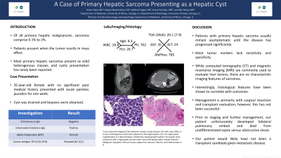
Case Description/Methods: A 35-year-old female presented with acute jaundice. Computed tomography (CT) of the abdomen revealed a large cystic lesion replacing most of the liver with associated mass effect causing biliary dilation (A). The CT also demonstrated adjacent heterogeneous masses within the anterior and posterior right hepatic lobe with regional lymphadenopathy (B). Laboratory evaluation revealed a white blood cell count of 20.6 K/uL, total bilirubin of 14.1 mg/dL, alkaline phosphatase of 785 U/L, normal AFP, and CA 19-9 of 61 U/mL. Echinococcus IgG was negative and Entamoeba histolytica IgG was positive after which the cyst was drained. Biopsy of the solid component revealed undifferentiated round cell sarcoma (C-D). Prior to staging and further management, the patient developed bilateral pulmonary emboli and died from undifferentiated septic or obstructive shock.
Discussion: Patients with primary hepatic sarcoma usually remain asymptomatic until the disease has progressed significantly. Most tumor markers lack sensitivity and specificity for sarcoma. While CT and magnetic resonance imaging (MRI) are commonly obtained in the evaluation of liver lesions, there are no characteristic imaging features for primary liver sarcoma due to its heterogeneous nature. Therefore, biopsy is required for the diagnosis. Our patient was ultimately diagnosed with undifferentiated round cell sarcoma. Interestingly, histological features of primary liver sarcoma have been shown to correlate with outcomes. This case highlights that despite it being exceedingly rare, primary hepatic sarcoma can present as a complex hepatic cyst and biopsy should be performed to rule out malignancy. Management of primary liver sarcoma is primarily with surgical resection. Patients with inoperable disease have poor outcomes due to early metastasis, primarily to the lungs. Liver transplantation has not been successful due to rapid tumor recurrence.

Disclosures:
Hasan Raza, MD1, Dexter E. Nwachukwu, DO, MS1, Michael Siegel, DO2, Grace Guzman, MD3, Jennifer Wang, MD1. P2506 - A Case of Primary Hepatic Sarcoma Presenting as a Hepatic Cyst, ACG 2023 Annual Scientific Meeting Abstracts. Vancouver, BC, Canada: American College of Gastroenterology.
1University of Illinois Chicago, Chicago, IL; 2University of Illinois-Chicago, Chicago, IL; 3University of Illinois at Chicago, College of Medicine, Chicago, IL
Introduction: Sarcomas are rare malignant tumors which arise from mesenchymal cells. Of all primary hepatic cancers, sarcomas comprise 0.1% to 2% of all hepatic cancers. Patients usually remain asymptomatic until the tumor grows to a clinically significant size. Most primary hepatic sarcomas present as solid heterogeneous masses, and cystic presentation of hepatic sarcoma has been rarely reported. Here, we present a rare case of primary hepatic sarcoma presenting as a hepatic cyst.
Case Description/Methods: A 35-year-old female presented with acute jaundice. Computed tomography (CT) of the abdomen revealed a large cystic lesion replacing most of the liver with associated mass effect causing biliary dilation (A). The CT also demonstrated adjacent heterogeneous masses within the anterior and posterior right hepatic lobe with regional lymphadenopathy (B). Laboratory evaluation revealed a white blood cell count of 20.6 K/uL, total bilirubin of 14.1 mg/dL, alkaline phosphatase of 785 U/L, normal AFP, and CA 19-9 of 61 U/mL. Echinococcus IgG was negative and Entamoeba histolytica IgG was positive after which the cyst was drained. Biopsy of the solid component revealed undifferentiated round cell sarcoma (C-D). Prior to staging and further management, the patient developed bilateral pulmonary emboli and died from undifferentiated septic or obstructive shock.
Discussion: Patients with primary hepatic sarcoma usually remain asymptomatic until the disease has progressed significantly. Most tumor markers lack sensitivity and specificity for sarcoma. While CT and magnetic resonance imaging (MRI) are commonly obtained in the evaluation of liver lesions, there are no characteristic imaging features for primary liver sarcoma due to its heterogeneous nature. Therefore, biopsy is required for the diagnosis. Our patient was ultimately diagnosed with undifferentiated round cell sarcoma. Interestingly, histological features of primary liver sarcoma have been shown to correlate with outcomes. This case highlights that despite it being exceedingly rare, primary hepatic sarcoma can present as a complex hepatic cyst and biopsy should be performed to rule out malignancy. Management of primary liver sarcoma is primarily with surgical resection. Patients with inoperable disease have poor outcomes due to early metastasis, primarily to the lungs. Liver transplantation has not been successful due to rapid tumor recurrence.

Figure: CT images of the abdomen reveal large hepatic cyst with mass effect in A and heterogeneous solid mass adjacent to large hepatic cyst in B. High power magnification in C demonstrates distinctive nested growth pattern of invasion most consistent with a high-grade sarcoma with round and pleomorphic cellular features. Malignant neoplastic cells are shown adjacent to necrosis, fibrosis, and inflammation in D.
Disclosures:
Hasan Raza indicated no relevant financial relationships.
Dexter Nwachukwu indicated no relevant financial relationships.
Michael Siegel indicated no relevant financial relationships.
Grace Guzman indicated no relevant financial relationships.
Jennifer Wang indicated no relevant financial relationships.
Hasan Raza, MD1, Dexter E. Nwachukwu, DO, MS1, Michael Siegel, DO2, Grace Guzman, MD3, Jennifer Wang, MD1. P2506 - A Case of Primary Hepatic Sarcoma Presenting as a Hepatic Cyst, ACG 2023 Annual Scientific Meeting Abstracts. Vancouver, BC, Canada: American College of Gastroenterology.
