Monday Poster Session
Category: Liver
P2541 - Exophytic Hepatic Hemangioma Masquerading as a Gastrointestinal Stromal Tumor
Monday, October 23, 2023
10:30 AM - 4:15 PM PT
Location: Exhibit Hall

Has Audio

Muhammad Farooq, MD
East Carolina University
Greenville, NC
Presenting Author(s)
Muhammad Farooq, MD1, Mohammad H. Farooq, MD2, Prashant Mudireddy, MD1
1East Carolina University, Greenville, NC; 2Shifa College of Medicine, Greenville, NC
Introduction: Hepatic Hemangiomas are the most frequently seen benign vascular liver masses(7-20%). They are usually located in the right lobe, < 3 cm in size, asymptomatic and often discovered incidentally. A rare subset called exophytic hemangiomas are a challenge to diagnose due to low sensitivity of radiological imaging and often atypical location of these lesions. We present a case of a large hepatic exophytic hemangioma that was initially thought to be a peri-gastric GIST.
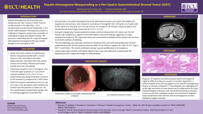
Case Description/Methods: 46 year old male with history of hypertension, obesity and gout presented with a 3 week history of dull, intermittent left sided abdominal pain associated with early satiety, nausea and vomiting. Physical examination and lab-work was unrevealing. Ultrasound showed a 11.8 cm echogenic mass in the left upper quadrant. CT scan with IV contrast revealed a 15.2 x 11.4 x 9.4 cm compressing mass along the greater curvature of the stomach (C) suspicious for GIST. EGD+EUS showed a well-defined 114 mm x 65 mm mass in the peri-gastric area(A+B). Fine needle biopsy showed bland spindle cells on cytology suggestive of possible GIST. Post procedure the patient developed sharp abdominal pain associated with diaphoresis, hypotension and syncope. Labs showed an acute drop in hemoglobin from 14.9g/dL to 10.6g/dL over 6 hours. CTA showed large peri-gastric hemorrhage measuring 16.4 x 9.5 x 11.5 cm. Emergent angiography showed nodular contrast enhancement of a lesion near the left hepatic lobe supplied by a segment of the left hepatic artery suggestive of a large exophytic hemangioma. The segmental artery was successfully embolized and the patient did not have any further evidence of bleeding. The final pathology was externally reviewed and confirmed proliferation of thick-walled blood vessels(D) with positive stains for SMA, CD 34, ERG but negative for S100, CD 117, Dog-1, GLUT 1 and Inhibin. The results confirmed a benign vascular proliferation or hemangioma. Laparoscopic exam showed a left lateral segment liver mass and underwent a laparoscopic left hepatectomy with surgical pathology currently pending.
Discussion: Diagnosis of exophytic hepatic hemangiomas can be challenging due to their often atypical location for a primary hepatic mass, non-specific heterogeneous appearance on contrast enhanced CT and vascularity of these lesions making tissue biopsy difficult. Histopathology with staining is more accurate than cytology and often important for diagnosis. Large symptomatic hemangiomas are best treated by surgical resection.

Disclosures:
Muhammad Farooq, MD1, Mohammad H. Farooq, MD2, Prashant Mudireddy, MD1. P2541 - Exophytic Hepatic Hemangioma Masquerading as a Gastrointestinal Stromal Tumor, ACG 2023 Annual Scientific Meeting Abstracts. Vancouver, BC, Canada: American College of Gastroenterology.
1East Carolina University, Greenville, NC; 2Shifa College of Medicine, Greenville, NC
Introduction: Hepatic Hemangiomas are the most frequently seen benign vascular liver masses(7-20%). They are usually located in the right lobe, < 3 cm in size, asymptomatic and often discovered incidentally. A rare subset called exophytic hemangiomas are a challenge to diagnose due to low sensitivity of radiological imaging and often atypical location of these lesions. We present a case of a large hepatic exophytic hemangioma that was initially thought to be a peri-gastric GIST.
Case Description/Methods: 46 year old male with history of hypertension, obesity and gout presented with a 3 week history of dull, intermittent left sided abdominal pain associated with early satiety, nausea and vomiting. Physical examination and lab-work was unrevealing. Ultrasound showed a 11.8 cm echogenic mass in the left upper quadrant. CT scan with IV contrast revealed a 15.2 x 11.4 x 9.4 cm compressing mass along the greater curvature of the stomach (C) suspicious for GIST. EGD+EUS showed a well-defined 114 mm x 65 mm mass in the peri-gastric area(A+B). Fine needle biopsy showed bland spindle cells on cytology suggestive of possible GIST. Post procedure the patient developed sharp abdominal pain associated with diaphoresis, hypotension and syncope. Labs showed an acute drop in hemoglobin from 14.9g/dL to 10.6g/dL over 6 hours. CTA showed large peri-gastric hemorrhage measuring 16.4 x 9.5 x 11.5 cm. Emergent angiography showed nodular contrast enhancement of a lesion near the left hepatic lobe supplied by a segment of the left hepatic artery suggestive of a large exophytic hemangioma. The segmental artery was successfully embolized and the patient did not have any further evidence of bleeding. The final pathology was externally reviewed and confirmed proliferation of thick-walled blood vessels(D) with positive stains for SMA, CD 34, ERG but negative for S100, CD 117, Dog-1, GLUT 1 and Inhibin. The results confirmed a benign vascular proliferation or hemangioma. Laparoscopic exam showed a left lateral segment liver mass and underwent a laparoscopic left hepatectomy with surgical pathology currently pending.
Discussion: Diagnosis of exophytic hepatic hemangiomas can be challenging due to their often atypical location for a primary hepatic mass, non-specific heterogeneous appearance on contrast enhanced CT and vascularity of these lesions making tissue biopsy difficult. Histopathology with staining is more accurate than cytology and often important for diagnosis. Large symptomatic hemangiomas are best treated by surgical resection.

Figure: A: Endoscopic view of perigastric mass causing gastric compression. B: Endosonographic view of perigastric mass.
C: CT image of perigastric mass. D: Histologic slide showing hemangioma with thick walled dilated vascular channels
C: CT image of perigastric mass. D: Histologic slide showing hemangioma with thick walled dilated vascular channels
Disclosures:
Muhammad Farooq indicated no relevant financial relationships.
Mohammad Farooq indicated no relevant financial relationships.
Prashant Mudireddy indicated no relevant financial relationships.
Muhammad Farooq, MD1, Mohammad H. Farooq, MD2, Prashant Mudireddy, MD1. P2541 - Exophytic Hepatic Hemangioma Masquerading as a Gastrointestinal Stromal Tumor, ACG 2023 Annual Scientific Meeting Abstracts. Vancouver, BC, Canada: American College of Gastroenterology.
