Monday Poster Session
Category: Liver
P2545 - Peliosis Hepatis Mimicking as Malignancy: A Case Report
Monday, October 23, 2023
10:30 AM - 4:15 PM PT
Location: Exhibit Hall

Has Audio
.jpg)
Mehwish Kishore, MD
UICOMP
Peoria, IL
Presenting Author(s)
Mehwish Kishore, MD1, Irfa Tariq, MBBS2, Lavneet Chawla, MD2, Saba Farooq, MD3, Muhammad Asghar, MD4, Imran Balouch, MD4
1UICOMP, Peoria, IL; 2OSF Saint Francis Medical Center, Peoria, IL; 3UIC Peoria, Peoria, IL; 4UIC, Peoria, IL
Introduction: Peliosis hepatis is a rare disease characterized by random proliferation of sinusoidal capillaries resulting in formation of blood-filled parenchymal cysts. Risk factors include exposure to certain drugs, immune disorders and infectious causes. We present a case of a patient who initially presented with cholangitis suspected to be secondary to common bile duct stone vs malignancy given multiple hepatic lesions. However, further workup revealed peliosis hepatis.
Case Description/Methods: A 64-year-old male presented with epigastric abdominal pain and jaundice. Lab data showed cholestatic transaminitis, as well as leukocytosis and thrombocytopenia. Blood cultures grew E Coli and K pneumoniae and IV antibiotics were initiated. Further workup including panel for chronic liver diseases and alpha-fetoprotein levels were unremarkable. CT abdomen revealed choledocholithiasis with multiple liver lesions and splenomegaly. Bone marrow biopsy with flow cytometry and pathology negative for malignancy. Patient received IVIg for suspected ITP. Due to suspected ascending cholangitis, endoscopic retrograde cholangiopancreatography (ERCP) was performed with stone removal and stent placement. Additionally, endoscopic ultrasound (EUS) with biopsy of one of the liver lesions was performed. Post op period was complicated by hemobilia and arteriobiliary fistula for which embolization of hepatic artery was performed. Hepatic lesion biopsy showed material consistent with cyst contents without evidence of malignancy. Based on the biopsy findings in conjunction with imaging and presentation, diagnosis of peliosis hepatis was made. With clearance of bacteremia and improvement in platelet counts, repeat MRI showed slight reduction in size of the hepatic lesions
Discussion: The pathogenesis of peliosis hepatis remains unclear but it may involve sinusoidal outflow obstruction leading to necrosis and subsequent endothelial dilation. Patients can be asymptomatic or present with abdominal pain, jaundice, portal hypertension or liver failure. CT angiography or MRI can identify hepatic lesions, but it is difficult to differentiate them from abscesses or metastases. Percutaneous biopsy can confirm the diagnosis but is associated with hemorrhage. There is no specific treatment for this condition, rather just treating the associated condition. Peliosis hepatis should be considered as a differential diagnosis when patients present with rapidly evolving hepatic lesions, especially in the setting of ITP and bacteremia, as in our case.

Disclosures:
Mehwish Kishore, MD1, Irfa Tariq, MBBS2, Lavneet Chawla, MD2, Saba Farooq, MD3, Muhammad Asghar, MD4, Imran Balouch, MD4. P2545 - Peliosis Hepatis Mimicking as Malignancy: A Case Report, ACG 2023 Annual Scientific Meeting Abstracts. Vancouver, BC, Canada: American College of Gastroenterology.
1UICOMP, Peoria, IL; 2OSF Saint Francis Medical Center, Peoria, IL; 3UIC Peoria, Peoria, IL; 4UIC, Peoria, IL
Introduction: Peliosis hepatis is a rare disease characterized by random proliferation of sinusoidal capillaries resulting in formation of blood-filled parenchymal cysts. Risk factors include exposure to certain drugs, immune disorders and infectious causes. We present a case of a patient who initially presented with cholangitis suspected to be secondary to common bile duct stone vs malignancy given multiple hepatic lesions. However, further workup revealed peliosis hepatis.
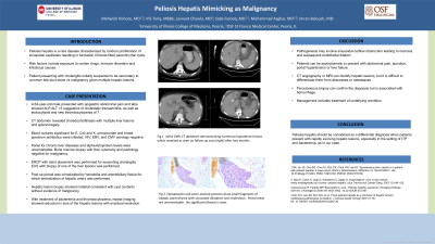
Case Description/Methods: A 64-year-old male presented with epigastric abdominal pain and jaundice. Lab data showed cholestatic transaminitis, as well as leukocytosis and thrombocytopenia. Blood cultures grew E Coli and K pneumoniae and IV antibiotics were initiated. Further workup including panel for chronic liver diseases and alpha-fetoprotein levels were unremarkable. CT abdomen revealed choledocholithiasis with multiple liver lesions and splenomegaly. Bone marrow biopsy with flow cytometry and pathology negative for malignancy. Patient received IVIg for suspected ITP. Due to suspected ascending cholangitis, endoscopic retrograde cholangiopancreatography (ERCP) was performed with stone removal and stent placement. Additionally, endoscopic ultrasound (EUS) with biopsy of one of the liver lesions was performed. Post op period was complicated by hemobilia and arteriobiliary fistula for which embolization of hepatic artery was performed. Hepatic lesion biopsy showed material consistent with cyst contents without evidence of malignancy. Based on the biopsy findings in conjunction with imaging and presentation, diagnosis of peliosis hepatis was made. With clearance of bacteremia and improvement in platelet counts, repeat MRI showed slight reduction in size of the hepatic lesions
Discussion: The pathogenesis of peliosis hepatis remains unclear but it may involve sinusoidal outflow obstruction leading to necrosis and subsequent endothelial dilation. Patients can be asymptomatic or present with abdominal pain, jaundice, portal hypertension or liver failure. CT angiography or MRI can identify hepatic lesions, but it is difficult to differentiate them from abscesses or metastases. Percutaneous biopsy can confirm the diagnosis but is associated with hemorrhage. There is no specific treatment for this condition, rather just treating the associated condition. Peliosis hepatis should be considered as a differential diagnosis when patients present with rapidly evolving hepatic lesions, especially in the setting of ITP and bacteremia, as in our case.

Figure: CT findings demonstrate multiple hypodense hepatic lesions varying in the size.
Disclosures:
Mehwish Kishore indicated no relevant financial relationships.
Irfa Tariq indicated no relevant financial relationships.
Lavneet Chawla indicated no relevant financial relationships.
Saba Farooq indicated no relevant financial relationships.
Muhammad Asghar indicated no relevant financial relationships.
Imran Balouch indicated no relevant financial relationships.
Mehwish Kishore, MD1, Irfa Tariq, MBBS2, Lavneet Chawla, MD2, Saba Farooq, MD3, Muhammad Asghar, MD4, Imran Balouch, MD4. P2545 - Peliosis Hepatis Mimicking as Malignancy: A Case Report, ACG 2023 Annual Scientific Meeting Abstracts. Vancouver, BC, Canada: American College of Gastroenterology.
