Monday Poster Session
Category: Liver
P2576 - An Uncommon Association: Cholangiocarcinoma and Autoimmune Hepatitis
Monday, October 23, 2023
10:30 AM - 4:15 PM PT
Location: Exhibit Hall

Has Audio

Kelli Kosako Yost, MD
University of Arizona College of Medicine
Phoenix, AZ
Presenting Author(s)
Kelli Kosako Yost, MD, Dayna Telken, DO, Zaid Alkaissy, MD, Natasha Narang, DO, Kevin Liu, MD, Karn Wijarnpreecha, MD, MPH, Ma Ai Thanda Han, MD
University of Arizona College of Medicine, Phoenix, AZ
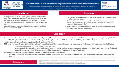
Introduction: Cholangiocarcinoma (CCA) is a rare type of epithelial cell tumor that arises in the intrahepatic, extrahepatic/distal, or perihilar biliary tree. Less than 10% of cases are intrahepatic. Risk factors of CCA include PSC, choledochal cysts, hepatolithiasis, viral hepatitis, and IBD. Expanding literature suggests that autoimmune hepatitis (AIH) may also be a risk factor for CCA.
Case Description/Methods: The patient is a 62 year old female with Hashimoto’s thyroiditis and recent diagnosis of biopsy-proven, ANA positive/SMA positive autoimmune hepatitis. On a steroid taper of 40 mg prednisone, she developed increased pruritis. Prednisone was increased to 60 mg daily, but subsequent lab work demonstrated ALT 1,633 U/L, AST 559 U/L, total bilirubin 32.1 mg/dL, alkaline phosphatase of 875 U/L, and CA 19-9 419 U/mL. Abdominal ultrasound demonstrated intra and extrahepatic ductal dilation without cholelithiasis. MRCP revealed intrahepatic biliary ductal dilation and a focus of restricted diffusion in the hepatic hilum. Repeat liver biopsy showed moderate cholestatic hepatitis with mild periportal fibrosis. ERCP showed severe common hepatic duct stricture with upstream dilatation of the intrahepatic ducts and visualization of irregular, ulcerated mucosa in the common hepatic bile duct. The stricture was dilated to 4mm, a plastic stent was placed, and biopsies/brushings were taken. Biopsies revealed atypical epithelial cells with nuclear overlapping, irregular nuclear membranes, and prominent nucleoli with wild-type staining of p53 and retained nuclear expression of SMAD4, equivocal for the presence of high grade malignancy, concerning for cholangiocarcinoma. FISH testing was unable to be performed due to insufficient specimen. Patient subsequently underwent extended right hepatectomy, cholecystectomy, and RNY hepaticojejunostomy. Intraoperative biopsy showed moderately differentiated intrahepatic cholangiocarcinoma at right tri-segment 5,6,7,8 and extrahepatic bile duct with perineural invasion.
Discussion: This is one of few case reports in the literature of development of CCA in the setting of AIH, in a patient with no other known risk factors. It is postulated that the chronic inflammatory response of AIH, specifically the infiltration of lymphocytes, plasma cells, and macrophages, creates a similar environment seen in other conditions predisposing to CCA. Due to this, special attention should be paid to this subset of patients in monitoring them for development of biliary malignancies.

Disclosures:
Kelli Kosako Yost, MD, Dayna Telken, DO, Zaid Alkaissy, MD, Natasha Narang, DO, Kevin Liu, MD, Karn Wijarnpreecha, MD, MPH, Ma Ai Thanda Han, MD. P2576 - An Uncommon Association: Cholangiocarcinoma and Autoimmune Hepatitis, ACG 2023 Annual Scientific Meeting Abstracts. Vancouver, BC, Canada: American College of Gastroenterology.
University of Arizona College of Medicine, Phoenix, AZ
Introduction: Cholangiocarcinoma (CCA) is a rare type of epithelial cell tumor that arises in the intrahepatic, extrahepatic/distal, or perihilar biliary tree. Less than 10% of cases are intrahepatic. Risk factors of CCA include PSC, choledochal cysts, hepatolithiasis, viral hepatitis, and IBD. Expanding literature suggests that autoimmune hepatitis (AIH) may also be a risk factor for CCA.
Case Description/Methods: The patient is a 62 year old female with Hashimoto’s thyroiditis and recent diagnosis of biopsy-proven, ANA positive/SMA positive autoimmune hepatitis. On a steroid taper of 40 mg prednisone, she developed increased pruritis. Prednisone was increased to 60 mg daily, but subsequent lab work demonstrated ALT 1,633 U/L, AST 559 U/L, total bilirubin 32.1 mg/dL, alkaline phosphatase of 875 U/L, and CA 19-9 419 U/mL. Abdominal ultrasound demonstrated intra and extrahepatic ductal dilation without cholelithiasis. MRCP revealed intrahepatic biliary ductal dilation and a focus of restricted diffusion in the hepatic hilum. Repeat liver biopsy showed moderate cholestatic hepatitis with mild periportal fibrosis. ERCP showed severe common hepatic duct stricture with upstream dilatation of the intrahepatic ducts and visualization of irregular, ulcerated mucosa in the common hepatic bile duct. The stricture was dilated to 4mm, a plastic stent was placed, and biopsies/brushings were taken. Biopsies revealed atypical epithelial cells with nuclear overlapping, irregular nuclear membranes, and prominent nucleoli with wild-type staining of p53 and retained nuclear expression of SMAD4, equivocal for the presence of high grade malignancy, concerning for cholangiocarcinoma. FISH testing was unable to be performed due to insufficient specimen. Patient subsequently underwent extended right hepatectomy, cholecystectomy, and RNY hepaticojejunostomy. Intraoperative biopsy showed moderately differentiated intrahepatic cholangiocarcinoma at right tri-segment 5,6,7,8 and extrahepatic bile duct with perineural invasion.
Discussion: This is one of few case reports in the literature of development of CCA in the setting of AIH, in a patient with no other known risk factors. It is postulated that the chronic inflammatory response of AIH, specifically the infiltration of lymphocytes, plasma cells, and macrophages, creates a similar environment seen in other conditions predisposing to CCA. Due to this, special attention should be paid to this subset of patients in monitoring them for development of biliary malignancies.

Figure: MRCP demonstrating tortuous dilation of the left and right hepatic ducts
Disclosures:
Kelli Kosako Yost indicated no relevant financial relationships.
Dayna Telken indicated no relevant financial relationships.
Zaid Alkaissy indicated no relevant financial relationships.
Natasha Narang indicated no relevant financial relationships.
Kevin Liu indicated no relevant financial relationships.
Karn Wijarnpreecha indicated no relevant financial relationships.
Ma Ai Thanda Han indicated no relevant financial relationships.
Kelli Kosako Yost, MD, Dayna Telken, DO, Zaid Alkaissy, MD, Natasha Narang, DO, Kevin Liu, MD, Karn Wijarnpreecha, MD, MPH, Ma Ai Thanda Han, MD. P2576 - An Uncommon Association: Cholangiocarcinoma and Autoimmune Hepatitis, ACG 2023 Annual Scientific Meeting Abstracts. Vancouver, BC, Canada: American College of Gastroenterology.
