Monday Poster Session
Category: Liver
P2579 - Cracking the Code: Molecular Analysis of Hepatic Adenomas
Monday, October 23, 2023
10:30 AM - 4:15 PM PT
Location: Exhibit Hall

Has Audio
- AP
Arsheya Patel, MD
The Ohio State University Wexner Medical Center
Columbus, OH
Presenting Author(s)
Arsheya Patel, MD1, Vivek Mendiratta, MD1, Chathur Acharya, MBBS2
1The Ohio State University Wexner Medical Center, Columbus, OH; 2Ohio State University, Columbus, OH
Introduction: Hepatocellular adenomas (HA) are benign monoclonal proliferations of hepatocytes that carry a potential for malignant transformation to hepatocellular carcinoma (HCC). HA demonstrate significant diversity at the molecular level resulting in distinct subtypes with variable risks of progression to HCC. Beta-catenin activated HCA result from a mutation in CTNNB1 and represent only 10% of all HA, but carry the highest risk of progression to HCC1,2. We describe a case of a young male who presented with a hepatic mass found to be HCC arising from a beta-catenin activated HA.
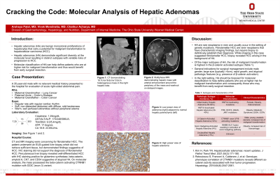
Case Description/Methods: A 35-year-old male presented for evaluation of right-sided abdominal pain and jaundice. He had no relevant past medical or family history. Contrast-enhanced CT demonstrated a 16.7cmx16.6cmx13.6cm heterogenous mass in the right lobe of the liver. Multiphase MRI showed arterial enhancement around the periphery of the mass with lack of central enhancement and washout on delayed images concerning for fibrolamellar HCC (FlHCC). EUS-guided fine needle aspiration revealed scant liver parenchyma with thickened hepatic plates and loss of reticulin staining suggestive of HCC. Immunohistochemical (IHC) staining did not support FIHCC. Subsequent percutaneous biopsy revealed well differentiated adenocarcinoma with IHC diffusely positive for glutamine synthetase, beta-catenin, amyloid A, CK7, and CD34 suggestive of atypical HA. On molecular analysis, the mass possessed the beta-catenin activating CTNNB1 mutation with S33C (exon 3) variant. Overall, the combination of IHC and molecular analyses suggested a well differentiated adenocarcinoma arising in the background of beta-catenin activated HA. After multidisciplinary discussion, the patient started medical treatment with immunotherapy.
Discussion: HA are rare neoplasms in men and usually occur in the setting of genetic mutations. FlHCC are rarer hepatic neoplasms that have a characteristic imaging finding, but require a liver biopsy for diagnosis. Though our patient had FlHCC on imaging, biopsy revealed instead adenocarcinoma arising from HA. Of the major subtypes of HA, beta-catenin activated HA carry the greatest risk of malignancy, particularly those with CTNNB1 exon 3 mutations as described in this case. In the right setting, HA should be biopsied for molecular classification to help define patients who are at higher risk for malignant transformation and consequently those who may benefit from early surgical resection.

Disclosures:
Arsheya Patel, MD1, Vivek Mendiratta, MD1, Chathur Acharya, MBBS2. P2579 - Cracking the Code: Molecular Analysis of Hepatic Adenomas, ACG 2023 Annual Scientific Meeting Abstracts. Vancouver, BC, Canada: American College of Gastroenterology.
1The Ohio State University Wexner Medical Center, Columbus, OH; 2Ohio State University, Columbus, OH
Introduction: Hepatocellular adenomas (HA) are benign monoclonal proliferations of hepatocytes that carry a potential for malignant transformation to hepatocellular carcinoma (HCC). HA demonstrate significant diversity at the molecular level resulting in distinct subtypes with variable risks of progression to HCC. Beta-catenin activated HCA result from a mutation in CTNNB1 and represent only 10% of all HA, but carry the highest risk of progression to HCC1,2. We describe a case of a young male who presented with a hepatic mass found to be HCC arising from a beta-catenin activated HA.
Case Description/Methods: A 35-year-old male presented for evaluation of right-sided abdominal pain and jaundice. He had no relevant past medical or family history. Contrast-enhanced CT demonstrated a 16.7cmx16.6cmx13.6cm heterogenous mass in the right lobe of the liver. Multiphase MRI showed arterial enhancement around the periphery of the mass with lack of central enhancement and washout on delayed images concerning for fibrolamellar HCC (FlHCC). EUS-guided fine needle aspiration revealed scant liver parenchyma with thickened hepatic plates and loss of reticulin staining suggestive of HCC. Immunohistochemical (IHC) staining did not support FIHCC. Subsequent percutaneous biopsy revealed well differentiated adenocarcinoma with IHC diffusely positive for glutamine synthetase, beta-catenin, amyloid A, CK7, and CD34 suggestive of atypical HA. On molecular analysis, the mass possessed the beta-catenin activating CTNNB1 mutation with S33C (exon 3) variant. Overall, the combination of IHC and molecular analyses suggested a well differentiated adenocarcinoma arising in the background of beta-catenin activated HA. After multidisciplinary discussion, the patient started medical treatment with immunotherapy.
Discussion: HA are rare neoplasms in men and usually occur in the setting of genetic mutations. FlHCC are rarer hepatic neoplasms that have a characteristic imaging finding, but require a liver biopsy for diagnosis. Though our patient had FlHCC on imaging, biopsy revealed instead adenocarcinoma arising from HA. Of the major subtypes of HA, beta-catenin activated HA carry the greatest risk of malignancy, particularly those with CTNNB1 exon 3 mutations as described in this case. In the right setting, HA should be biopsied for molecular classification to help define patients who are at higher risk for malignant transformation and consequently those who may benefit from early surgical resection.

Figure: T2-weighted MRI of Well Differentiated Adenocarcinoma of the Liver
Disclosures:
Arsheya Patel indicated no relevant financial relationships.
Vivek Mendiratta indicated no relevant financial relationships.
Chathur Acharya indicated no relevant financial relationships.
Arsheya Patel, MD1, Vivek Mendiratta, MD1, Chathur Acharya, MBBS2. P2579 - Cracking the Code: Molecular Analysis of Hepatic Adenomas, ACG 2023 Annual Scientific Meeting Abstracts. Vancouver, BC, Canada: American College of Gastroenterology.
