Monday Poster Session
Category: Liver
P2584 - Hepatic Angiomyolipoma
Monday, October 23, 2023
10:30 AM - 4:15 PM PT
Location: Exhibit Hall

Has Audio

Sumedha Singh, MD
Einstein Medical Center
Philadelphia, PA
Presenting Author(s)
Sumedha Singh, MD, Richard Kalman, MD, Maria Lagarde, MD
Einstein Medical Center, Philadelphia, PA
Introduction: We present a case of an hepatic angiomyolipoma. Angiomyolipomas are a rare, benign tumor composed of smooth muscle cells, adipose tissue, and vessels. Angiomyolipomas are most commonly found in the kidney, however can also be found in other organs including the liver, and are considered to be part of the perivascular epithelioid cell tumor (PEComa) family.
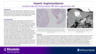
Case Description/Methods: 56F with history of hypercalcuria, nephrolithiasis, and duplicated left renal collecting system presented for finding of hepatic lobe lesion of 3cm on ultrasound performed for intermittent RUQ abdominal discomfort. Dedicated triple phase CT imaging showed a 4.2 x 3.8 x 4.0 cm hypervascular, arterially enhancing mass in segment 8 extending to segment 4A/B with an additional 1.2 x 1.2 cm centrally located lesion, concerning for malignancy initially thought to be due to fibrolamellar hepatocellular carcinoma. AFP 8.9. CA19-9 < 2. CEA 1.9, and all other bloodwork also normal. Her oncologic family history is remarkable for metastatic bladder cancer in father, osteocolesteoblastoma in the mouth in mother, maternal grandfather died from lymphoma. Patient is up to date on age-appropriate cancer screenings and with no known baseline liver disease. She underwent liver biopsy of the lesion with pathology showing rare hepatic finding of angiomyolipoma, with histological immunostains positive for HMB-45 and Melan-A. She was seen by surgery, interventional radiology, hepatology, oncology and genetics. The plan was ultimately made for local regional treatment with embolization and ablation by IR. However, the patient opted to defer treatment and continue close surveillance with imaging. Patient intends to complete genetic testing for NF1, TSC1, TSC2, VHL for this finding of angiomyolipoma due to the association with tuberous sclerosis syndromes.
Discussion: Angiomyolipomas are a rare, typically benign mesenchymal tumor that most often affect the kidneys but can affect other organs including the liver. Hepatic angiomyolipomas are rare but are in the differential for liver lesions in a patient without chronic liver disease and tissue sampling should be considered if the diagnosis is obscure based on radiographic appearance. The appearance can be suggestive of HCC due to non-specific radiographic findings and thus tissue diagnosis is essential. Management of this tumor varies ranging from surveillance to surgical resection.

Disclosures:
Sumedha Singh, MD, Richard Kalman, MD, Maria Lagarde, MD. P2584 - Hepatic Angiomyolipoma, ACG 2023 Annual Scientific Meeting Abstracts. Vancouver, BC, Canada: American College of Gastroenterology.
Einstein Medical Center, Philadelphia, PA
Introduction: We present a case of an hepatic angiomyolipoma. Angiomyolipomas are a rare, benign tumor composed of smooth muscle cells, adipose tissue, and vessels. Angiomyolipomas are most commonly found in the kidney, however can also be found in other organs including the liver, and are considered to be part of the perivascular epithelioid cell tumor (PEComa) family.
Case Description/Methods: 56F with history of hypercalcuria, nephrolithiasis, and duplicated left renal collecting system presented for finding of hepatic lobe lesion of 3cm on ultrasound performed for intermittent RUQ abdominal discomfort. Dedicated triple phase CT imaging showed a 4.2 x 3.8 x 4.0 cm hypervascular, arterially enhancing mass in segment 8 extending to segment 4A/B with an additional 1.2 x 1.2 cm centrally located lesion, concerning for malignancy initially thought to be due to fibrolamellar hepatocellular carcinoma. AFP 8.9. CA19-9 < 2. CEA 1.9, and all other bloodwork also normal. Her oncologic family history is remarkable for metastatic bladder cancer in father, osteocolesteoblastoma in the mouth in mother, maternal grandfather died from lymphoma. Patient is up to date on age-appropriate cancer screenings and with no known baseline liver disease. She underwent liver biopsy of the lesion with pathology showing rare hepatic finding of angiomyolipoma, with histological immunostains positive for HMB-45 and Melan-A. She was seen by surgery, interventional radiology, hepatology, oncology and genetics. The plan was ultimately made for local regional treatment with embolization and ablation by IR. However, the patient opted to defer treatment and continue close surveillance with imaging. Patient intends to complete genetic testing for NF1, TSC1, TSC2, VHL for this finding of angiomyolipoma due to the association with tuberous sclerosis syndromes.
Discussion: Angiomyolipomas are a rare, typically benign mesenchymal tumor that most often affect the kidneys but can affect other organs including the liver. Hepatic angiomyolipomas are rare but are in the differential for liver lesions in a patient without chronic liver disease and tissue sampling should be considered if the diagnosis is obscure based on radiographic appearance. The appearance can be suggestive of HCC due to non-specific radiographic findings and thus tissue diagnosis is essential. Management of this tumor varies ranging from surveillance to surgical resection.

Figure: Cytomorphologic features compatible with PECOMA (perivascular epithelioid cell neoplasm), most likely representing a component of an angiomyolipoma. The specimen consists of atypical cells with vacuolated cytoplasm, scattered blood vessels and some background liver parenchyma. Immunostains were performed to further characterize the atypical cells. The cells are positive for HMB-45 and Melan-A. Immunostain for CD34 highlights the vascular network.
Disclosures:
Sumedha Singh indicated no relevant financial relationships.
Richard Kalman indicated no relevant financial relationships.
Maria Lagarde indicated no relevant financial relationships.
Sumedha Singh, MD, Richard Kalman, MD, Maria Lagarde, MD. P2584 - Hepatic Angiomyolipoma, ACG 2023 Annual Scientific Meeting Abstracts. Vancouver, BC, Canada: American College of Gastroenterology.
