Monday Poster Session
Category: Liver
P2594 - Recanalization of the Paraumbilical Vein: A Unique Collateral Pathway in Decompensated Liver Cirrhosis
Monday, October 23, 2023
10:30 AM - 4:15 PM PT
Location: Exhibit Hall

Has Audio

Naga Venkata Rama Krishna Vura, MD
UTHSCSA
San Antonio, TX
Presenting Author(s)
Abu baker Khan, MBBS1, Pir Ahmad Shah, MD2, Naga Venkata Rama Krishna Vura, MD3
1Khyber Medical University, Peshawar, Northern Areas, Pakistan; 2University of Texas Health Science Center at San Antonio, San Antonio, TX; 3UTHSCSA, San Antonio, TX
Introduction: Portal hypertension is a common complication of cirrhosis, that often leads to redirect blood away from the liver and the development of collateral vessels as an adaptive response. We report a very interesting radiological finding of recanalization of the paraumbilical vein, serving as an additional collateral pathway in a patient with decompensated cirrhosis.
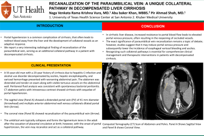
Case Description/Methods: A 55-year-old man with a 23-year history of cirrhosis due to hepatitis C infection and alcohol use disorder decompensated by ascites, hepatic encephalopathy, and variceal hemorrhage presented with worsening abdominal pain. The abdomen was distended and tender on exam along with visible tortuous vessels on the abdominal wall. Peritoneal fluid analysis was consistent with spontaneous bacterial peritonitis. CT abdomen pelvis with intravenous contrast showed cirrhosis with sequelae of portal hypertension. The sagittal view (Panel A) showed a distended portal vein (PV) of 41 mm diameter (Arrowhead) and multiple anterior abdominal wall venous collaterals dilated portal Vein (Arrow). The coronal view (Panel B) showed recanalization of the paraumbilical vein (Arrow). The umbilical vein typically collapses and forms the ligamentum teres in the adult after the cessation of placental circulation at birth; however, with the onset of portal hypertension, the vein may recanalize and act as a collateral pathway.
Discussion: In cirrhotic liver disease, increased resistance to portal blood flow leads to elevated portal venous pressure, often resulting in the reopening of occluded vessels. The exact significance of paraumbilical vein recanalization remains a topic of debate; however, studies suggest that it may reduce portal venous pressure and subsequently lower the incidence of esophageal variceal bleeding and ascites. Recognizing such collateral pathways is essential for comprehensive clinical management and therapeutic interventions in patients with decompensated cirrhosis.

Disclosures:
Abu baker Khan, MBBS1, Pir Ahmad Shah, MD2, Naga Venkata Rama Krishna Vura, MD3. P2594 - Recanalization of the Paraumbilical Vein: A Unique Collateral Pathway in Decompensated Liver Cirrhosis, ACG 2023 Annual Scientific Meeting Abstracts. Vancouver, BC, Canada: American College of Gastroenterology.
1Khyber Medical University, Peshawar, Northern Areas, Pakistan; 2University of Texas Health Science Center at San Antonio, San Antonio, TX; 3UTHSCSA, San Antonio, TX
Introduction: Portal hypertension is a common complication of cirrhosis, that often leads to redirect blood away from the liver and the development of collateral vessels as an adaptive response. We report a very interesting radiological finding of recanalization of the paraumbilical vein, serving as an additional collateral pathway in a patient with decompensated cirrhosis.
Case Description/Methods: A 55-year-old man with a 23-year history of cirrhosis due to hepatitis C infection and alcohol use disorder decompensated by ascites, hepatic encephalopathy, and variceal hemorrhage presented with worsening abdominal pain. The abdomen was distended and tender on exam along with visible tortuous vessels on the abdominal wall. Peritoneal fluid analysis was consistent with spontaneous bacterial peritonitis. CT abdomen pelvis with intravenous contrast showed cirrhosis with sequelae of portal hypertension. The sagittal view (Panel A) showed a distended portal vein (PV) of 41 mm diameter (Arrowhead) and multiple anterior abdominal wall venous collaterals dilated portal Vein (Arrow). The coronal view (Panel B) showed recanalization of the paraumbilical vein (Arrow). The umbilical vein typically collapses and forms the ligamentum teres in the adult after the cessation of placental circulation at birth; however, with the onset of portal hypertension, the vein may recanalize and act as a collateral pathway.
Discussion: In cirrhotic liver disease, increased resistance to portal blood flow leads to elevated portal venous pressure, often resulting in the reopening of occluded vessels. The exact significance of paraumbilical vein recanalization remains a topic of debate; however, studies suggest that it may reduce portal venous pressure and subsequently lower the incidence of esophageal variceal bleeding and ascites. Recognizing such collateral pathways is essential for comprehensive clinical management and therapeutic interventions in patients with decompensated cirrhosis.

Figure: Computed Tomography (CT) Scan of Abdomen and Pelvis. Panel A Shows Sagittal View and Panel B shows Coronal View
Disclosures:
Abu baker Khan indicated no relevant financial relationships.
Pir Ahmad Shah indicated no relevant financial relationships.
Naga Venkata Rama Krishna Vura indicated no relevant financial relationships.
Abu baker Khan, MBBS1, Pir Ahmad Shah, MD2, Naga Venkata Rama Krishna Vura, MD3. P2594 - Recanalization of the Paraumbilical Vein: A Unique Collateral Pathway in Decompensated Liver Cirrhosis, ACG 2023 Annual Scientific Meeting Abstracts. Vancouver, BC, Canada: American College of Gastroenterology.
