Monday Poster Session
Category: Small Intestine
P2647 - Encapsulating Sclerosing Peritonitis: A Rare Cause of Gastrointestinal Obstruction
Monday, October 23, 2023
10:30 AM - 4:15 PM PT
Location: Exhibit Hall

Has Audio

Seema Mir, MD
University of South Alabama Health Systems
Mobile, AL
Presenting Author(s)
Seema Mir, MD1, Anas Khouri, MD2, Xuebao Zhang, MD1, Abhijin Das, MD1, Benjamin Niland, MD1
1University of South Alabama Health Systems, Mobile, AL; 2University of South Alabama, Mobile, AL
Introduction: Encapsulating sclerosing peritonitis (ESP) is a rare complication of peritoneal dialysis (PD) that is characterized by intraperitoneal inflammation and fibrosis which can result in the encasement of bowel loops. It has a high morbidity and mortality rate and can lead to ultrafiltration failure and bowel obstruction.
Case Description/Methods: A 48-year-old African American woman presented with intractable nausea and vomiting, abdominal pain, and unintentional 30-pound weight loss for 6 months. Past medical history was significant for end-stage renal disease due to hypertension. Hemodialysis (HD) was her initial modality until she received a deceased donor renal transplant. She had chronic allograft nephropathy leading to graft failure 5 years since transplant and subsequently she was placed on PD for 7 years before transitioning to home HD.
On initial evaluation, CT Abdomen showed large volume abdominal and pelvic ascites, partially calcified lesion abutting the liver, and a calcification near the confluence of the right hepatic duct. She underwent a paracentesis with removal of bloody ascitic fluid, with a low serum ascites albumin gradient and negative cytology for malignancy. The patient's malignancy and infectious workup was negative.
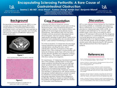
On readmission, CT Abdomen illustrated increased size of the partially calcified lesion in the right upper quadrant abutting the liver and the right lower quadrant lesion near the pelvic sidewall (Figure 1). Surgery was consulted for diagnostic laparotomy and peritoneal biopsies. The peritoneum was abnormally thickened, tanned and leathery in appearance with dense adhesion and grossly matted abdominal content covered by thickened visceral peritoneum layer. The pathology reported nodular fibrocollagenous proliferation (Figure 2). Based on these findings, the patient was diagnosed with ESP and started on Tamoxifen and Prednisone.
Discussion: ESP is a rare disease characterized by the intestines becoming partially or completely encased by a fibrous membrane. It is a complication of PD with prevalence varying between 0.4% to 8.9%, an incidence rate between 0.7 to 13.6 per 1,000 patient-years, and a risk of occurrence after 5 years on PD between 0.6% and 6.6%. The diagnosis of ESP is based on a combination of structural and functional features and is confirmed radiographically or by laparotomy. It is important to be clinically aware of the possibility of ESP for several years after discontinuing PD. ESP presents after withdrawal from PD in the majority of patients.

Disclosures:
Seema Mir, MD1, Anas Khouri, MD2, Xuebao Zhang, MD1, Abhijin Das, MD1, Benjamin Niland, MD1. P2647 - Encapsulating Sclerosing Peritonitis: A Rare Cause of Gastrointestinal Obstruction, ACG 2023 Annual Scientific Meeting Abstracts. Vancouver, BC, Canada: American College of Gastroenterology.
1University of South Alabama Health Systems, Mobile, AL; 2University of South Alabama, Mobile, AL
Introduction: Encapsulating sclerosing peritonitis (ESP) is a rare complication of peritoneal dialysis (PD) that is characterized by intraperitoneal inflammation and fibrosis which can result in the encasement of bowel loops. It has a high morbidity and mortality rate and can lead to ultrafiltration failure and bowel obstruction.
Case Description/Methods: A 48-year-old African American woman presented with intractable nausea and vomiting, abdominal pain, and unintentional 30-pound weight loss for 6 months. Past medical history was significant for end-stage renal disease due to hypertension. Hemodialysis (HD) was her initial modality until she received a deceased donor renal transplant. She had chronic allograft nephropathy leading to graft failure 5 years since transplant and subsequently she was placed on PD for 7 years before transitioning to home HD.
On initial evaluation, CT Abdomen showed large volume abdominal and pelvic ascites, partially calcified lesion abutting the liver, and a calcification near the confluence of the right hepatic duct. She underwent a paracentesis with removal of bloody ascitic fluid, with a low serum ascites albumin gradient and negative cytology for malignancy. The patient's malignancy and infectious workup was negative.
On readmission, CT Abdomen illustrated increased size of the partially calcified lesion in the right upper quadrant abutting the liver and the right lower quadrant lesion near the pelvic sidewall (Figure 1). Surgery was consulted for diagnostic laparotomy and peritoneal biopsies. The peritoneum was abnormally thickened, tanned and leathery in appearance with dense adhesion and grossly matted abdominal content covered by thickened visceral peritoneum layer. The pathology reported nodular fibrocollagenous proliferation (Figure 2). Based on these findings, the patient was diagnosed with ESP and started on Tamoxifen and Prednisone.
Discussion: ESP is a rare disease characterized by the intestines becoming partially or completely encased by a fibrous membrane. It is a complication of PD with prevalence varying between 0.4% to 8.9%, an incidence rate between 0.7 to 13.6 per 1,000 patient-years, and a risk of occurrence after 5 years on PD between 0.6% and 6.6%. The diagnosis of ESP is based on a combination of structural and functional features and is confirmed radiographically or by laparotomy. It is important to be clinically aware of the possibility of ESP for several years after discontinuing PD. ESP presents after withdrawal from PD in the majority of patients.

Figure: Figure 1. RLQ calcified lesion near the pelvic sidewall.
Figure 2. Nodular fibrocollagenous proliferation of peritoneum seen on pathology.
Figure 2. Nodular fibrocollagenous proliferation of peritoneum seen on pathology.
Disclosures:
Seema Mir indicated no relevant financial relationships.
Anas Khouri indicated no relevant financial relationships.
Xuebao Zhang indicated no relevant financial relationships.
Abhijin Das indicated no relevant financial relationships.
Benjamin Niland indicated no relevant financial relationships.
Seema Mir, MD1, Anas Khouri, MD2, Xuebao Zhang, MD1, Abhijin Das, MD1, Benjamin Niland, MD1. P2647 - Encapsulating Sclerosing Peritonitis: A Rare Cause of Gastrointestinal Obstruction, ACG 2023 Annual Scientific Meeting Abstracts. Vancouver, BC, Canada: American College of Gastroenterology.
