Monday Poster Session
Category: Small Intestine
P2681 - A Rare Case of IgG4-Related Sclerosing Mesenteritis Mimicking Small Bowel Malignancy!
Monday, October 23, 2023
10:30 AM - 4:15 PM PT
Location: Exhibit Hall

Has Audio

Ruchir Paladiya, MBBS
UConn Health
Hartford, CT
Presenting Author(s)
Ruchir Paladiya, MBBS1, Neil Khoury, MD2, Stephen Simeone, MD3, Jason Jacob, MD4, John Birk, MD, FACG5
1UConn Health, Hartford, CT; 2UConn Health, West Hartford, CT; 3University of Connecticut Health Center, Hartford, CT; 4Hartford Hospital, Hartford, CT; 5University of Connecticut Health Center, Farmington, CT
Introduction: IgG4-related sclerosing mesenteritis (IgG4-SM) is a rare, insidiously progressive fibroinflammatory disorder involving the adipose tissue of the small bowel mesentery. It is usually asymptomatic; however, extreme presentations include bowel obstruction, obstructive uropathy, chylous ascites and mesenteric ischemia. Mimickers include malignant lymphoma, desmoid tumors, carcinoid tumors, and panniculitis.
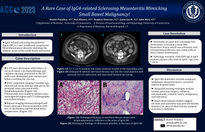
Case Description/Methods: A 69-year-old woman with history of cervical cancer status post chemotherapy, and radiation therapy, presented to the ED with acute abdominal pain, nausea, and bilious vomiting for one day. On admission, she was afebrile, tachycardic (115/min), normotensive. Her abdomen was soft, non-distended, with generalized tenderness. She denied eating spoiled food or past surgical history. Her infectious work-up was unremarkable. Prior outpatient imaging five months ago showed soft tissue density adjacent to the proximal colon associated with lymphadenopathy. A colonoscopy was attempted to assess for colonic involvement, but scope could not be passed beyond the rectosigmoid region and CT colonography was indeterminate. In the ED, the CT scan showed the increased size of the mass to 2.6 x 1.8cm with terminal ileum compression, causing ileal narrowing and proximal GI dilation. She was made NPO, and a nasogastric tube was placed for gastric decompression. Due to concern for malignancy and inability to reach the affected area endoscopically, she underwent open ileo-colectomy. A mass-like mesenteric lesion was extracted and retroperitoneal fibrosis was noted. There was no evidence of colonic involvement. She underwent cystectomy due to significant bladder adhesions. Pathology showed diffuse storiform sclerosis mixed with lymphoplasmacytic infiltrates, staining >100/HPF IgG & IgG4 positive plasma cells, with an IgG4: IgG ratio > 40%, negative for malignancy and obliterative phlebitis (Figure A-B).
Discussion: An association between IgG4-SM and retroperitoneal fibrosis has been described in the literature. Suspected etiologies include trauma, previous surgery, radiation, autoimmunity, ischemic injury, malignancy, and infections. The Patient’s radiation history along with histopathological, immunohistochemical & imaging findings suggested the diagnosis of IgG4-SM, a subset of sclerosing mesenteritis. There is some data from small observational studies which suggest that steroids and tamoxifen may provide benefit, however, in cases of obstructions, surgical resection is definitive.

Disclosures:
Ruchir Paladiya, MBBS1, Neil Khoury, MD2, Stephen Simeone, MD3, Jason Jacob, MD4, John Birk, MD, FACG5. P2681 - A Rare Case of IgG4-Related Sclerosing Mesenteritis Mimicking Small Bowel Malignancy!, ACG 2023 Annual Scientific Meeting Abstracts. Vancouver, BC, Canada: American College of Gastroenterology.
1UConn Health, Hartford, CT; 2UConn Health, West Hartford, CT; 3University of Connecticut Health Center, Hartford, CT; 4Hartford Hospital, Hartford, CT; 5University of Connecticut Health Center, Farmington, CT
Introduction: IgG4-related sclerosing mesenteritis (IgG4-SM) is a rare, insidiously progressive fibroinflammatory disorder involving the adipose tissue of the small bowel mesentery. It is usually asymptomatic; however, extreme presentations include bowel obstruction, obstructive uropathy, chylous ascites and mesenteric ischemia. Mimickers include malignant lymphoma, desmoid tumors, carcinoid tumors, and panniculitis.
Case Description/Methods: A 69-year-old woman with history of cervical cancer status post chemotherapy, and radiation therapy, presented to the ED with acute abdominal pain, nausea, and bilious vomiting for one day. On admission, she was afebrile, tachycardic (115/min), normotensive. Her abdomen was soft, non-distended, with generalized tenderness. She denied eating spoiled food or past surgical history. Her infectious work-up was unremarkable. Prior outpatient imaging five months ago showed soft tissue density adjacent to the proximal colon associated with lymphadenopathy. A colonoscopy was attempted to assess for colonic involvement, but scope could not be passed beyond the rectosigmoid region and CT colonography was indeterminate. In the ED, the CT scan showed the increased size of the mass to 2.6 x 1.8cm with terminal ileum compression, causing ileal narrowing and proximal GI dilation. She was made NPO, and a nasogastric tube was placed for gastric decompression. Due to concern for malignancy and inability to reach the affected area endoscopically, she underwent open ileo-colectomy. A mass-like mesenteric lesion was extracted and retroperitoneal fibrosis was noted. There was no evidence of colonic involvement. She underwent cystectomy due to significant bladder adhesions. Pathology showed diffuse storiform sclerosis mixed with lymphoplasmacytic infiltrates, staining >100/HPF IgG & IgG4 positive plasma cells, with an IgG4: IgG ratio > 40%, negative for malignancy and obliterative phlebitis (Figure A-B).
Discussion: An association between IgG4-SM and retroperitoneal fibrosis has been described in the literature. Suspected etiologies include trauma, previous surgery, radiation, autoimmunity, ischemic injury, malignancy, and infections. The Patient’s radiation history along with histopathological, immunohistochemical & imaging findings suggested the diagnosis of IgG4-SM, a subset of sclerosing mesenteritis. There is some data from small observational studies which suggest that steroids and tamoxifen may provide benefit, however, in cases of obstructions, surgical resection is definitive.

Figure: Figure A: Histological findings of storiform fibrosis in the mass of IgG4-SM
Figure B: Histological findings of obliterative phlebitis in the mass of IgG4-SM
Figure B: Histological findings of obliterative phlebitis in the mass of IgG4-SM
Disclosures:
Ruchir Paladiya indicated no relevant financial relationships.
Neil Khoury indicated no relevant financial relationships.
Stephen Simeone indicated no relevant financial relationships.
Jason Jacob indicated no relevant financial relationships.
John Birk indicated no relevant financial relationships.
Ruchir Paladiya, MBBS1, Neil Khoury, MD2, Stephen Simeone, MD3, Jason Jacob, MD4, John Birk, MD, FACG5. P2681 - A Rare Case of IgG4-Related Sclerosing Mesenteritis Mimicking Small Bowel Malignancy!, ACG 2023 Annual Scientific Meeting Abstracts. Vancouver, BC, Canada: American College of Gastroenterology.
