Monday Poster Session
Category: Small Intestine
P2693 - A Rare Case of Duodenal Intussusception Caused by Migrated Percutaneous Endoscopic Gastrostomy Feeding Tube
Monday, October 23, 2023
10:30 AM - 4:15 PM PT
Location: Exhibit Hall

Has Audio
- AE
Adnan Elghezewi, MD
Marshall University Joan C. Edwards School of Medicine
Huntington, WV
Presenting Author(s)
Abdelwahap Elghezewi, MD1, Mohamed Hammad, MD1, Mujtaba Mohamed, MBBS2, Adnan Elghezewi, MD3, Hafiz Zarsham Ali Ikram, MD4, Wesam Frandah, MD3
1Marshall University, Huntington, WV; 2Joan C. Edwards School of Medicine, Huntington, WV; 3Marshall University Joan C. Edwards School of Medicine, Huntington, WV; 4Marshall University School of Medicine, Huntington, WV
Introduction: Intussusception is invagination of the intestine. It is a rare finding in adults, representing only 5% of all cases of intussusception. Duodenal intussusception is also rare since duodenum is fixed in retroperitoneum . It is caused by benign or malignant tumors of the duodenum, malrotation abnormalities, and Meckel’s diverticulum Is almost never reported as A case of duodenal intussusception due to migrated feeding tube. Herein, we present a case of adult duodeno-duodenal intussusception due to migrated Percutaneous Gastrostomy tube.
Case Description/Methods: 50-year-old white lady with a past medical history of TBI complicated by quadriplegia, spasticity, dysphagia status post-PEG tube, and seizure disorder. presented to the emergency department for nausea, vomiting, and abdominal distention. vital signs heart rate: 128 BPM, Blood pressure 125/90, respiratory rate 20, Oxygen saturation 96% on room air, weight 40.9 kg. Contractures were noted on both lower limbs. An abdominal exam revealed a distended abdomen with a mild leak around the PEG tube. Laboratory data: White cell counts 38 , hemoglobin 13.1 , platelets 308 , MCV 86. fl , sodium 131 , creatinine 1.2 , procalcitonin 5.08. lactic acid 4.6 mmol. CT scan of the abdomen showed dilated duodenum (6 cm), and intussusception within the duodenum and volvulus in the same location. The stomach was distended and full of fluids. There was dilation of intra and extra-hepatic biliary tracts. A feeding tube was pulled back and placed with gravity. Around 1100 ml of bilious fluid drained. Abdominal distention improved. A repeat CT scan showed improvement in duodenal intussusception and volvulus. On the following day, the patient had upper endoscopy which showed intact gastrostomy with a patent G-tube present in the gastric body. Two non-bleeding linear duodenal ulcers with no stigmata of bleeding 2nd and 3rd part of the duodenum with no evidence of stenosis, mass, or deformity in the entire duodenum. The patient was discharged back to the nursing home two days later.
Discussion: Duodenal intussusception in adults is a rare condition with nonspecific clinical manifestation. It can present with acute, subacute or intermittent symptoms. It is most common in middle-aged women. CT abdomen is the most accurate test and upper endoscopy is needed to rule out any intraluminal causes. Treatment of choice is either endoscopic resection or surgical resection depending on the size and depth of extension of the lesion into the luminal wall.

Disclosures:
Abdelwahap Elghezewi, MD1, Mohamed Hammad, MD1, Mujtaba Mohamed, MBBS2, Adnan Elghezewi, MD3, Hafiz Zarsham Ali Ikram, MD4, Wesam Frandah, MD3. P2693 - A Rare Case of Duodenal Intussusception Caused by Migrated Percutaneous Endoscopic Gastrostomy Feeding Tube, ACG 2023 Annual Scientific Meeting Abstracts. Vancouver, BC, Canada: American College of Gastroenterology.
1Marshall University, Huntington, WV; 2Joan C. Edwards School of Medicine, Huntington, WV; 3Marshall University Joan C. Edwards School of Medicine, Huntington, WV; 4Marshall University School of Medicine, Huntington, WV
Introduction: Intussusception is invagination of the intestine. It is a rare finding in adults, representing only 5% of all cases of intussusception. Duodenal intussusception is also rare since duodenum is fixed in retroperitoneum . It is caused by benign or malignant tumors of the duodenum, malrotation abnormalities, and Meckel’s diverticulum Is almost never reported as A case of duodenal intussusception due to migrated feeding tube. Herein, we present a case of adult duodeno-duodenal intussusception due to migrated Percutaneous Gastrostomy tube.
Case Description/Methods: 50-year-old white lady with a past medical history of TBI complicated by quadriplegia, spasticity, dysphagia status post-PEG tube, and seizure disorder. presented to the emergency department for nausea, vomiting, and abdominal distention. vital signs heart rate: 128 BPM, Blood pressure 125/90, respiratory rate 20, Oxygen saturation 96% on room air, weight 40.9 kg. Contractures were noted on both lower limbs. An abdominal exam revealed a distended abdomen with a mild leak around the PEG tube. Laboratory data: White cell counts 38 , hemoglobin 13.1 , platelets 308 , MCV 86. fl , sodium 131 , creatinine 1.2 , procalcitonin 5.08. lactic acid 4.6 mmol. CT scan of the abdomen showed dilated duodenum (6 cm), and intussusception within the duodenum and volvulus in the same location. The stomach was distended and full of fluids. There was dilation of intra and extra-hepatic biliary tracts. A feeding tube was pulled back and placed with gravity. Around 1100 ml of bilious fluid drained. Abdominal distention improved. A repeat CT scan showed improvement in duodenal intussusception and volvulus. On the following day, the patient had upper endoscopy which showed intact gastrostomy with a patent G-tube present in the gastric body. Two non-bleeding linear duodenal ulcers with no stigmata of bleeding 2nd and 3rd part of the duodenum with no evidence of stenosis, mass, or deformity in the entire duodenum. The patient was discharged back to the nursing home two days later.
Discussion: Duodenal intussusception in adults is a rare condition with nonspecific clinical manifestation. It can present with acute, subacute or intermittent symptoms. It is most common in middle-aged women. CT abdomen is the most accurate test and upper endoscopy is needed to rule out any intraluminal causes. Treatment of choice is either endoscopic resection or surgical resection depending on the size and depth of extension of the lesion into the luminal wall.

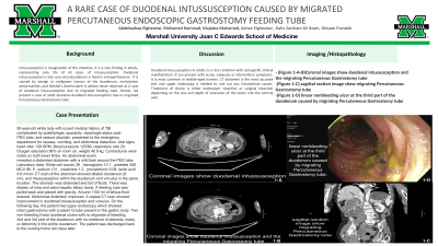
Figure: -Coronal images show duodenal intussusception and the migrating Percutaneous Gastrostomy tube
-sagittal section image show migrating Percutaneous Gastrostomy tube
-linear nonbleeding ulcer at the third part of the duodenum caused by migrating Percutaneous Gastrostomy tube
-sagittal section image show migrating Percutaneous Gastrostomy tube
-linear nonbleeding ulcer at the third part of the duodenum caused by migrating Percutaneous Gastrostomy tube
Disclosures:
Abdelwahap Elghezewi indicated no relevant financial relationships.
Mohamed Hammad indicated no relevant financial relationships.
Mujtaba Mohamed indicated no relevant financial relationships.
Adnan Elghezewi indicated no relevant financial relationships.
Hafiz Zarsham Ali Ikram indicated no relevant financial relationships.
Wesam Frandah: Endo gastric solutions – Consultant. Olympus Medical – Consultant.
Abdelwahap Elghezewi, MD1, Mohamed Hammad, MD1, Mujtaba Mohamed, MBBS2, Adnan Elghezewi, MD3, Hafiz Zarsham Ali Ikram, MD4, Wesam Frandah, MD3. P2693 - A Rare Case of Duodenal Intussusception Caused by Migrated Percutaneous Endoscopic Gastrostomy Feeding Tube, ACG 2023 Annual Scientific Meeting Abstracts. Vancouver, BC, Canada: American College of Gastroenterology.
