Monday Poster Session
Category: Small Intestine
P2694 - Primary Squamous Cell Carcinoma of the Small Bowel
Monday, October 23, 2023
10:30 AM - 4:15 PM PT
Location: Exhibit Hall

Has Audio

Amir Riaz, MD
Larkin Community Hospital
Miami, FL
Presenting Author(s)
Amir Riaz, MD1, Zaid Rana, DO2, Ana Hernandez. Caballero, MD3, Karthik Mohan, DO1
1Larkin Community Hospital, Miami, FL; 2Larkin Community Hospital, South Miami, FL; 3North Shore Medical Center, Miami, FL
Introduction: Primary squamous cell carcinoma (SCC) of the small bowel is extremely rare, with a global incidence of < 1 per 100,000 population. The pathogenesis is unclear given that the small bowel does not normally contain squamous cells. Clinical symptoms may be similar to other tumors of the ileum. Given its rarity and unclear pathophysiology, it is important for caregivers to take notice of this rare and possible diagnosis.
Case Description/Methods: A 52-year old African American male presented to the Emergency Department for abdominal distention, nausea and non-bilious, non-bloody vomiting. Medical history was notable for alcohol-related cirrhosis, melanoma of the skin, and hypertension. Physical exam was notable for a tympanic and distended abdomen. Laboratory studies showed WBC 4.4 k/ul, Hgb 10.5 g/dl, MCV 92 fL, Na 135 mmol/L, and K 3.3 mmol/L. Non contrast enhanced computed tomography (CT) scan of the abdomen and pelvis revealed high-grade small bowel obstruction with abrupt transition in the right abdomen. At the site of the transition there was a 4.5 cm length of small bowel which demonstrated circumferential mural thickening and luminal narrowing, highly concerning for small bowel malignant neoplasm. The patient refused decompressive methods, thus underwent exploratory laparotomy which revealed an obstructing mass in the distal ileum. He subsequently underwent small-bowel resection. Pathology from the mass was notable for moderately differentiated squamous cell carcinoma. Mass was 5 x 4.2 x 1.2 cm, well circumscribed, tan-pink fungating, ulcerated mass.
Discussion: Compared to other gastrointestinal organs, malignant tumors of the small intestine are very rare. A better understanding of the pathogenesis and clinical features of this rare and malignant tumor is necessary to make an early diagnosis and optimize treatment. It is often difficult to delineate whether SCC is primary or metastatic. The most frequent tumors metastasizing to the small intestine include, carcinomas of the lung, breast, kidneys, colon, and melanoma. Imaging can help identify if the tumor originated somewhere else. Early detection may improve prognosis and prevent complications such as perforation. Due to the scarcity of data available in the literature, the effects of chemotherapy and radiotherapy on the survival and recurrence are unknown.

Disclosures:
Amir Riaz, MD1, Zaid Rana, DO2, Ana Hernandez. Caballero, MD3, Karthik Mohan, DO1. P2694 - Primary Squamous Cell Carcinoma of the Small Bowel, ACG 2023 Annual Scientific Meeting Abstracts. Vancouver, BC, Canada: American College of Gastroenterology.
1Larkin Community Hospital, Miami, FL; 2Larkin Community Hospital, South Miami, FL; 3North Shore Medical Center, Miami, FL
Introduction: Primary squamous cell carcinoma (SCC) of the small bowel is extremely rare, with a global incidence of < 1 per 100,000 population. The pathogenesis is unclear given that the small bowel does not normally contain squamous cells. Clinical symptoms may be similar to other tumors of the ileum. Given its rarity and unclear pathophysiology, it is important for caregivers to take notice of this rare and possible diagnosis.
Case Description/Methods: A 52-year old African American male presented to the Emergency Department for abdominal distention, nausea and non-bilious, non-bloody vomiting. Medical history was notable for alcohol-related cirrhosis, melanoma of the skin, and hypertension. Physical exam was notable for a tympanic and distended abdomen. Laboratory studies showed WBC 4.4 k/ul, Hgb 10.5 g/dl, MCV 92 fL, Na 135 mmol/L, and K 3.3 mmol/L. Non contrast enhanced computed tomography (CT) scan of the abdomen and pelvis revealed high-grade small bowel obstruction with abrupt transition in the right abdomen. At the site of the transition there was a 4.5 cm length of small bowel which demonstrated circumferential mural thickening and luminal narrowing, highly concerning for small bowel malignant neoplasm. The patient refused decompressive methods, thus underwent exploratory laparotomy which revealed an obstructing mass in the distal ileum. He subsequently underwent small-bowel resection. Pathology from the mass was notable for moderately differentiated squamous cell carcinoma. Mass was 5 x 4.2 x 1.2 cm, well circumscribed, tan-pink fungating, ulcerated mass.
Discussion: Compared to other gastrointestinal organs, malignant tumors of the small intestine are very rare. A better understanding of the pathogenesis and clinical features of this rare and malignant tumor is necessary to make an early diagnosis and optimize treatment. It is often difficult to delineate whether SCC is primary or metastatic. The most frequent tumors metastasizing to the small intestine include, carcinomas of the lung, breast, kidneys, colon, and melanoma. Imaging can help identify if the tumor originated somewhere else. Early detection may improve prognosis and prevent complications such as perforation. Due to the scarcity of data available in the literature, the effects of chemotherapy and radiotherapy on the survival and recurrence are unknown.

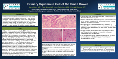
Figure: A. Hematoxylin and Eosin stain, at 100x magnification, showing the tumor interface, at the top there is residual small bowel glandular epithelium overlying nests of squamous cell carcinoma.
B. Hematoxylin and Eosin stain, at 200x magnification, showing squamous cell carcinoma with areas of keratinization (keratin pearls).
C. Hematoxylin and Eosin stain, at 600x magnification, showing the intercellular bridges (desmosomes) between the epithelial cells of squamous cell carcinoma.
B. Hematoxylin and Eosin stain, at 200x magnification, showing squamous cell carcinoma with areas of keratinization (keratin pearls).
C. Hematoxylin and Eosin stain, at 600x magnification, showing the intercellular bridges (desmosomes) between the epithelial cells of squamous cell carcinoma.
Disclosures:
Amir Riaz indicated no relevant financial relationships.
Zaid Rana indicated no relevant financial relationships.
Ana Caballero indicated no relevant financial relationships.
Karthik Mohan indicated no relevant financial relationships.
Amir Riaz, MD1, Zaid Rana, DO2, Ana Hernandez. Caballero, MD3, Karthik Mohan, DO1. P2694 - Primary Squamous Cell Carcinoma of the Small Bowel, ACG 2023 Annual Scientific Meeting Abstracts. Vancouver, BC, Canada: American College of Gastroenterology.
