Monday Poster Session
Category: Small Intestine
P2700 - A Case of Jejunal Metastatic Malignant Melanoma Presenting as Hematochezia: An Unusual Cause of Gastrointestinal Bleed
Monday, October 23, 2023
10:30 AM - 4:15 PM PT
Location: Exhibit Hall

Has Audio

Balakrishna Ravella, MBBS, MD
OSF Health St. Mary's Medical Center
Galesburg, IL
Presenting Author(s)
Balakrishna Ravella, MBBS, MD1, Roy Kondapavuluru, MBBS, MD2, Nestor Pamatmat, MD1, Mikala Brinkman, MD1, Thomas Whittle, MD1, Afshin Khaiser, MD1
1OSF Health St. Mary's Medical Center, Galesburg, IL; 2Knapp Medical Center, Weslaco, TX
Introduction: Malignant melanoma (MM) is the fifth most common cancer in the United States. Widespread metastasis remains the main cause of death. While the most common metastatic sites being the lung, liver and central nervous system; metastasis to the small bowel is relatively uncommon. We present a patient with history of MM who presented with hematochezia and was found to have a jejunal metastatic melanoma.
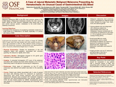
Case Description/Methods: A 52-year-old male with a history of stage IB T2N0M0 malignant melanoma of upper back that was treated with wide surgical excision with sentinel lymph node biopsy 10 years ago, presented to our emergency department with abdominal cramping, hematochezia and lightheadedness for 2 days and a 15-pound unintentional weight loss in 3 weeks. Patient was found to have a hemoglobin of 6.0 gm/dl, dropped from a baseline hemoglobin of 15.0 gm/dl. A computed tomography (CT) scan of the abdomen and pelvis with contrast was done which showed a 3.9 x 5.3 cm obstructing mass in the jejunum concerning for malignancy (Figure 1&2). Given signs of obstruction on the CT scan, endoscopy was deferred, and he was taken to the operating room, after resuscitation with 2 units of packed red blood cells. Patient underwent an exploratory laparotomy with small bowel resection (Figure 3&4) and end-to-end-anastomosis. Biopsies were consistent with metastatic malignant melanoma and immunohistochemistry was strongly positive for S100 (Figure 5&6). A BRAF mutation assay was positive for V600E. The patient is currently undergoing treatment with a combination of ipilimumab and nivolumab.
Discussion: Malignant melanoma most commonly tends to metastasize to regional lymph nodes followed by lungs, liver, brain, bone and adrenal gland. Among patients who are diagnosed with cutaneous melanoma, approximately 50% have a GI metastasis, however symptomatic GI involvement of melanoma is only seen in 1-4% of patients and is a frequent autopsy finding (60%). The most common location of small bowel melanoma is the terminal ileum followed by jejunum and duodenum. Imaging studies such as CT abdomen and pelvis play a vital role in diagnosing GI melanomas, as endoscopic examination of the small bowel is limited. In majority of reported cases, diagnosis was relied on radiologic exam and confirmation by exploratory laparotomy.
In conclusion, metastatic melanoma of the small bowel should be suspected in any patient with a previous history of MM and presenting with a GI bleed; even though the presentation is after several years.

Disclosures:
Balakrishna Ravella, MBBS, MD1, Roy Kondapavuluru, MBBS, MD2, Nestor Pamatmat, MD1, Mikala Brinkman, MD1, Thomas Whittle, MD1, Afshin Khaiser, MD1. P2700 - A Case of Jejunal Metastatic Malignant Melanoma Presenting as Hematochezia: An Unusual Cause of Gastrointestinal Bleed, ACG 2023 Annual Scientific Meeting Abstracts. Vancouver, BC, Canada: American College of Gastroenterology.
1OSF Health St. Mary's Medical Center, Galesburg, IL; 2Knapp Medical Center, Weslaco, TX
Introduction: Malignant melanoma (MM) is the fifth most common cancer in the United States. Widespread metastasis remains the main cause of death. While the most common metastatic sites being the lung, liver and central nervous system; metastasis to the small bowel is relatively uncommon. We present a patient with history of MM who presented with hematochezia and was found to have a jejunal metastatic melanoma.
Case Description/Methods: A 52-year-old male with a history of stage IB T2N0M0 malignant melanoma of upper back that was treated with wide surgical excision with sentinel lymph node biopsy 10 years ago, presented to our emergency department with abdominal cramping, hematochezia and lightheadedness for 2 days and a 15-pound unintentional weight loss in 3 weeks. Patient was found to have a hemoglobin of 6.0 gm/dl, dropped from a baseline hemoglobin of 15.0 gm/dl. A computed tomography (CT) scan of the abdomen and pelvis with contrast was done which showed a 3.9 x 5.3 cm obstructing mass in the jejunum concerning for malignancy (Figure 1&2). Given signs of obstruction on the CT scan, endoscopy was deferred, and he was taken to the operating room, after resuscitation with 2 units of packed red blood cells. Patient underwent an exploratory laparotomy with small bowel resection (Figure 3&4) and end-to-end-anastomosis. Biopsies were consistent with metastatic malignant melanoma and immunohistochemistry was strongly positive for S100 (Figure 5&6). A BRAF mutation assay was positive for V600E. The patient is currently undergoing treatment with a combination of ipilimumab and nivolumab.
Discussion: Malignant melanoma most commonly tends to metastasize to regional lymph nodes followed by lungs, liver, brain, bone and adrenal gland. Among patients who are diagnosed with cutaneous melanoma, approximately 50% have a GI metastasis, however symptomatic GI involvement of melanoma is only seen in 1-4% of patients and is a frequent autopsy finding (60%). The most common location of small bowel melanoma is the terminal ileum followed by jejunum and duodenum. Imaging studies such as CT abdomen and pelvis play a vital role in diagnosing GI melanomas, as endoscopic examination of the small bowel is limited. In majority of reported cases, diagnosis was relied on radiologic exam and confirmation by exploratory laparotomy.
In conclusion, metastatic melanoma of the small bowel should be suspected in any patient with a previous history of MM and presenting with a GI bleed; even though the presentation is after several years.

Figure: 1. Axial view of the CT scan of abdomen and pelvis showing the small bowel mass
2. Coronal view of the CT scan of abdomen and pelvis showing the small bowel mass and dilated upstream bowel loops with multiple air fluid levels
3. Serosal surface of the jejunum showing round, whitish tumor mass
4. Jejunal mucosa showing large necrotic tan yellow tumor mass extending through the jejunal wall without any gross perforation
5. Tumor cells showing epithelioid shape, hyperchromasia and intranuclear inclusions consistent with melanoma
6. Tumor cells staining positive for S-100 immunohistochemistry stain
Disclosures:
Balakrishna Ravella indicated no relevant financial relationships.
Roy Kondapavuluru indicated no relevant financial relationships.
Nestor Pamatmat indicated no relevant financial relationships.
Mikala Brinkman indicated no relevant financial relationships.
Thomas Whittle indicated no relevant financial relationships.
Afshin Khaiser indicated no relevant financial relationships.
Balakrishna Ravella, MBBS, MD1, Roy Kondapavuluru, MBBS, MD2, Nestor Pamatmat, MD1, Mikala Brinkman, MD1, Thomas Whittle, MD1, Afshin Khaiser, MD1. P2700 - A Case of Jejunal Metastatic Malignant Melanoma Presenting as Hematochezia: An Unusual Cause of Gastrointestinal Bleed, ACG 2023 Annual Scientific Meeting Abstracts. Vancouver, BC, Canada: American College of Gastroenterology.
