Monday Poster Session
Category: Stomach
P2780 - Gastric Pseudomelanosis: An Uncommon Endoscopic Finding
Monday, October 23, 2023
10:30 AM - 4:15 PM PT
Location: Exhibit Hall

Has Audio

Seema Mir, MD
University of South Alabama Health Systems
Mobile, AL
Presenting Author(s)
Seema Mir, MD1, Elizabeth Craig, DO2, Mary C. Marshall, MD3
1University of South Alabama Health Systems, Mobile, AL; 2DO, Mobile, AL; 3USA Health University Hospital, Mobile, AL
Introduction: Pseudomelanoiss duodeni has been reported previously in literature, however pseudomelanosis gastri remains a rare endoscopic finding. The dark brown or black speckling appearance of the mucosa is due to deposition of iron sulfide pigments in the lamina propria of the mucosal cells. Here we discuss an 80-year-old patient with a history of end stage renal disease (ESRD) presenting with melena and anemia found to have esophageal and gastric pseudomelanosis.
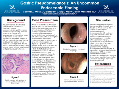
Case Description/Methods: An 80-year-old African American female with past medical history of ESRD on hemodialysis, anemia of chronic disease, atrial fibrillation on Eliquis, hypertension and peripheral vascular disease presented with an occlusion of her left arteriovenous fistula. During her hospitalization, the patient developed anemia with an acute drop in her hemoglobin from 8 g/dl to 6 g/dl and several melenic stools. She underwent an Esophagogastroduodenoscopy which illustrated abnormal blackened mucosa of the distal esophagus (Figure 1) and abnormal mucosa in the body, prepyloric and incisura of the stomach with blackened spots and evidence of inflammatory mucosa (Figure 2). Biopsies were taken, with pathology showing columnar epithelium with extensive intestinal metaplasia and chronic active gastritis with intestinal metaplasia. Immunostaining was negative for Helicobacter pylori. There was no evidence of melanosis coli on colonoscopy.
Discussion: Pseudomelanosis is an rare and benign condition typically seen in the 5th to 7th decade of life. Gastric and duodenal pseudomelanosis is inconsequential with no known long term sequelae. There are several possible etiologies of pseudomelanosis, including chronic oral iron therapy, hypertension, antihypertensive medications, ESRD, diabetes, hemochromatosis and gastrointestinal bleeding. Pathogenesis remains unclear. In our patient with anemia of chronic disease and ESRD, a possible explanation would be inflammation induced IL-6 resulting in increased hepcidin transcription ultimately resulting in increased iron storage in cells.

Disclosures:
Seema Mir, MD1, Elizabeth Craig, DO2, Mary C. Marshall, MD3. P2780 - Gastric Pseudomelanosis: An Uncommon Endoscopic Finding, ACG 2023 Annual Scientific Meeting Abstracts. Vancouver, BC, Canada: American College of Gastroenterology.
1University of South Alabama Health Systems, Mobile, AL; 2DO, Mobile, AL; 3USA Health University Hospital, Mobile, AL
Introduction: Pseudomelanoiss duodeni has been reported previously in literature, however pseudomelanosis gastri remains a rare endoscopic finding. The dark brown or black speckling appearance of the mucosa is due to deposition of iron sulfide pigments in the lamina propria of the mucosal cells. Here we discuss an 80-year-old patient with a history of end stage renal disease (ESRD) presenting with melena and anemia found to have esophageal and gastric pseudomelanosis.
Case Description/Methods: An 80-year-old African American female with past medical history of ESRD on hemodialysis, anemia of chronic disease, atrial fibrillation on Eliquis, hypertension and peripheral vascular disease presented with an occlusion of her left arteriovenous fistula. During her hospitalization, the patient developed anemia with an acute drop in her hemoglobin from 8 g/dl to 6 g/dl and several melenic stools. She underwent an Esophagogastroduodenoscopy which illustrated abnormal blackened mucosa of the distal esophagus (Figure 1) and abnormal mucosa in the body, prepyloric and incisura of the stomach with blackened spots and evidence of inflammatory mucosa (Figure 2). Biopsies were taken, with pathology showing columnar epithelium with extensive intestinal metaplasia and chronic active gastritis with intestinal metaplasia. Immunostaining was negative for Helicobacter pylori. There was no evidence of melanosis coli on colonoscopy.
Discussion: Pseudomelanosis is an rare and benign condition typically seen in the 5th to 7th decade of life. Gastric and duodenal pseudomelanosis is inconsequential with no known long term sequelae. There are several possible etiologies of pseudomelanosis, including chronic oral iron therapy, hypertension, antihypertensive medications, ESRD, diabetes, hemochromatosis and gastrointestinal bleeding. Pathogenesis remains unclear. In our patient with anemia of chronic disease and ESRD, a possible explanation would be inflammation induced IL-6 resulting in increased hepcidin transcription ultimately resulting in increased iron storage in cells.

Figure: Figure 1: Black pigment seen in the distal esophagus
Figure 2: Abnormal mucosa and black pigmentation of the stomach
Figure 2: Abnormal mucosa and black pigmentation of the stomach
Disclosures:
Seema Mir indicated no relevant financial relationships.
Elizabeth Craig indicated no relevant financial relationships.
Mary Marshall indicated no relevant financial relationships.
Seema Mir, MD1, Elizabeth Craig, DO2, Mary C. Marshall, MD3. P2780 - Gastric Pseudomelanosis: An Uncommon Endoscopic Finding, ACG 2023 Annual Scientific Meeting Abstracts. Vancouver, BC, Canada: American College of Gastroenterology.
