Monday Poster Session
Category: Stomach
P2781 - When One Hernia Is Not Enough: A Patient With a Large Type-III Hiatal Hernia and Ventral Wall Hernia
Monday, October 23, 2023
10:30 AM - 4:15 PM PT
Location: Exhibit Hall

Has Audio
- TW
TJ L. Wilson, MD
Detroit Medical Center/Wayne State University
Detroit, MI
Presenting Author(s)
TJ L. Wilson, MD1, Sophia Haroon Dar, MD2, Nooraldin Merza, MD3, Yousaf Zafar, MD4, Rehmat Ullah Awan, MD5, Amna Iqbal, MD3, Mohamad I.. Itani, MD1
1Detroit Medical Center/Wayne State University, Detroit, MI; 2Northshore-Long Island Jewish Medical Center, New York, NY; 3University of Toledo, Toledo, OH; 4University of Mississippi Medical Center, Jackson, MS; 5Ochsner Rush Medical Center, Meridian, MS
Introduction: Type-III hiatal hernias account for approximately 5% of hiatal hernia and often present with symptoms consistent with GERD. Initial management is with lifestyle modification and proton pump inhibitors (PPIs); for more severe symptoms, endoscopic or surgical hernia repair remain an option. Ventral hernias are also common in adults and vary in symptomatology depending on the size of the defect and presence of complications such as incarceration and strangulation. We present a patient a large type-III hiatal and an incarcerated ventral wall hernia who was managed successfully with surgical repair.
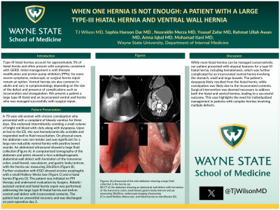
Case Description/Methods: A 70-year-old woman with chronic constipation who presented with a complaint of bloody vomitus for three days. She endorsed intermittently vomiting a small volume of bright red blood with clots along with dyspepsia. Upon arrival to the ED, she was hemodynamically unstable and responded well to fluid resuscitation. On physical exam, her abdomen was non-tender and was significant for a large non-reducible ventral hernia with positive bowel sounds. An abdominal ultrasound showed a large fluid collection (Figure A). A computerized tomography of the abdomen and pelvis showed a 4cm subdiaphragmatic abdominal wall defect with herniation of the transverse colon, small bowel, vasculature, and gastric body/antrum, with the hernia sac measuring 20x20cm (Figure B). Further evaluation with EGD showed erosive esophagitis with a small Mallory-Weiss tear (Figure C) and a hiatal hernia (Figures D). The patient was initiated on PPI therapy and underwent evaluation by Surgery. Robotic-assisted ventral and hiatal hernia repair was performed, addressing the large type-III hiatal hernia and 6x4cm ventral wall defect with incarcerated contents. The patient had an uneventful recovery and was discharged on post-operative day 3.
Discussion: While most hiatal hernias can be managed conservatively, our patient presented with atypical features for a type-III hiatal hernia, including hematemesis, which was further complicated by an incarcerated ventral hernia involving the stomach, small and large bowels. The patient's dyspepsia likely resulted from the hiatal hernia, while constipation was likely due to the incarcerated contents. Surgical intervention was deemed necessary to address both the hiatal and ventral hernias, leading to a successful outcome. This case highlights the need for individualized management in patients with complex hernias involving multiple defects.

Disclosures:
TJ L. Wilson, MD1, Sophia Haroon Dar, MD2, Nooraldin Merza, MD3, Yousaf Zafar, MD4, Rehmat Ullah Awan, MD5, Amna Iqbal, MD3, Mohamad I.. Itani, MD1. P2781 - When One Hernia Is Not Enough: A Patient With a Large Type-III Hiatal Hernia and Ventral Wall Hernia, ACG 2023 Annual Scientific Meeting Abstracts. Vancouver, BC, Canada: American College of Gastroenterology.
1Detroit Medical Center/Wayne State University, Detroit, MI; 2Northshore-Long Island Jewish Medical Center, New York, NY; 3University of Toledo, Toledo, OH; 4University of Mississippi Medical Center, Jackson, MS; 5Ochsner Rush Medical Center, Meridian, MS
Introduction: Type-III hiatal hernias account for approximately 5% of hiatal hernia and often present with symptoms consistent with GERD. Initial management is with lifestyle modification and proton pump inhibitors (PPIs); for more severe symptoms, endoscopic or surgical hernia repair remain an option. Ventral hernias are also common in adults and vary in symptomatology depending on the size of the defect and presence of complications such as incarceration and strangulation. We present a patient a large type-III hiatal and an incarcerated ventral wall hernia who was managed successfully with surgical repair.
Case Description/Methods: A 70-year-old woman with chronic constipation who presented with a complaint of bloody vomitus for three days. She endorsed intermittently vomiting a small volume of bright red blood with clots along with dyspepsia. Upon arrival to the ED, she was hemodynamically unstable and responded well to fluid resuscitation. On physical exam, her abdomen was non-tender and was significant for a large non-reducible ventral hernia with positive bowel sounds. An abdominal ultrasound showed a large fluid collection (Figure A). A computerized tomography of the abdomen and pelvis showed a 4cm subdiaphragmatic abdominal wall defect with herniation of the transverse colon, small bowel, vasculature, and gastric body/antrum, with the hernia sac measuring 20x20cm (Figure B). Further evaluation with EGD showed erosive esophagitis with a small Mallory-Weiss tear (Figure C) and a hiatal hernia (Figures D). The patient was initiated on PPI therapy and underwent evaluation by Surgery. Robotic-assisted ventral and hiatal hernia repair was performed, addressing the large type-III hiatal hernia and 6x4cm ventral wall defect with incarcerated contents. The patient had an uneventful recovery and was discharged on post-operative day 3.
Discussion: While most hiatal hernias can be managed conservatively, our patient presented with atypical features for a type-III hiatal hernia, including hematemesis, which was further complicated by an incarcerated ventral hernia involving the stomach, small and large bowels. The patient's dyspepsia likely resulted from the hiatal hernia, while constipation was likely due to the incarcerated contents. Surgical intervention was deemed necessary to address both the hiatal and ventral hernias, leading to a successful outcome. This case highlights the need for individualized management in patients with complex hernias involving multiple defects.

Figure: Figures: (A) ultrasound of the mid-abdomen showing a large fluid collection in the hernia sac, (B) CT of the abdomen showing an abdominal wall defect with herniation of the transverse colon, small bowel, gastric body/antrum and sac measuring 20x20cm, endoscopic imaging showcasing (C) a small Mallory-Weiss tear, and hiatal hernia on retroflexion (D).
Disclosures:
TJ Wilson indicated no relevant financial relationships.
Sophia Haroon Dar indicated no relevant financial relationships.
Nooraldin Merza indicated no relevant financial relationships.
Yousaf Zafar indicated no relevant financial relationships.
Rehmat Ullah Awan indicated no relevant financial relationships.
Amna Iqbal indicated no relevant financial relationships.
Mohamad Itani indicated no relevant financial relationships.
TJ L. Wilson, MD1, Sophia Haroon Dar, MD2, Nooraldin Merza, MD3, Yousaf Zafar, MD4, Rehmat Ullah Awan, MD5, Amna Iqbal, MD3, Mohamad I.. Itani, MD1. P2781 - When One Hernia Is Not Enough: A Patient With a Large Type-III Hiatal Hernia and Ventral Wall Hernia, ACG 2023 Annual Scientific Meeting Abstracts. Vancouver, BC, Canada: American College of Gastroenterology.
