Monday Poster Session
Category: Stomach
P2797 - Acute Gastric Volvulus: A Case Against Borchardt’s Triad With the Presence of Cameron Lesions
Monday, October 23, 2023
10:30 AM - 4:15 PM PT
Location: Exhibit Hall

Has Audio
- AA
Adnan Ansari, DO
Franciscan Health
Olympia Fields, IL
Presenting Author(s)
Adnan Ansari, DO1, Rahil Desai, DO2, Varshita Goduguchinta, DO3, Nicholas Baur, DO3, Roy Putnam, DO4, Ashirf Al-Ghanoudi, MD, MPH, FACG4
1Franciscan Health, Olympia Fields, IL; 2Franciscan Health Olympia Fields, Westlake, OH; 3Franciscan Health Olympia Fields, Chicago, IL; 4Franciscan Health Olympia Fields, Olympia Fields, IL
Introduction: Acute gastric volvulus is a rare but potentially deadly condition that has a mortality rate between 30 to 50%, prompting early diagnosis and intervention. It often presents with a trio of symptoms known as Borchardt’s triad: severe epigastric pain, intractable retching without vomiting, and inability to pass an NG tube. However, in 25% of cases, these symptoms may not be obvious, and are often misdiagnosed. Patients with large gastric volvulus can also present with Cameron lesions, which are large linear ulcerations on gastric mucosal folds. The overall presence of Cameron lesions found on EGDs are less than 1%. We highlight a unique case of gastric volvulus due to a large hiatal hernia with the presence of Cameron lesions on EGD that did not present with the typical Borchardt’s triad.
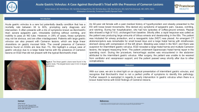
Case Description/Methods: An 82-year old female with a past medical history of hypothyroidism and obesity presented to the ED with loose bowel movements. She denied any symptoms of epigastric pain, nausea, vomiting, and retching. During her hospitalization, she had two episodes of coffee-ground emesis. Repeat labs showed a Hgb of 10.5, unchanged from baseline. Shortly after, a rapid response was called as the patient was producing large amounts of bilious emesis and desaturating in the 80s. The patient was intubated for airway protection, and a nasogastric tube (NGT) was placed. An emergent CT abdomen/pelvis was remarkable for small bowel ileus and a large hiatal hernia with intrathoracic stomach along with compression of the left atrium. Bleeding was noted in the NGT, and there was suspicion for intermittent gastric volvulus. EGD revealed a large hiatal hernia and multiple Cameron lesions, the largest measuring 5mm. The patient underwent laparoscopic hiatal hernia repair in the operating room. During the procedure, hemorrhagic ascites was encountered in the abdomen attributed to the intermittent gastric volvulus. After surgery, the patient was unable to be weaned from ventilator and vasopressor support, and the patient passed away shortly after due to other complications.
Discussion: With our case, we aim to shed light on an atypical presentation of intermittent gastric volvulus, and recognize that Borchardt’s triad is not a perfect profile of symptoms to identify this pathology. Further research is warranted in regards to early intervention in gastric volvulus when there is a known hiatal hernia with EGD findings of Cameron lesions.

Disclosures:
Adnan Ansari, DO1, Rahil Desai, DO2, Varshita Goduguchinta, DO3, Nicholas Baur, DO3, Roy Putnam, DO4, Ashirf Al-Ghanoudi, MD, MPH, FACG4. P2797 - Acute Gastric Volvulus: A Case Against Borchardt’s Triad With the Presence of Cameron Lesions, ACG 2023 Annual Scientific Meeting Abstracts. Vancouver, BC, Canada: American College of Gastroenterology.
1Franciscan Health, Olympia Fields, IL; 2Franciscan Health Olympia Fields, Westlake, OH; 3Franciscan Health Olympia Fields, Chicago, IL; 4Franciscan Health Olympia Fields, Olympia Fields, IL
Introduction: Acute gastric volvulus is a rare but potentially deadly condition that has a mortality rate between 30 to 50%, prompting early diagnosis and intervention. It often presents with a trio of symptoms known as Borchardt’s triad: severe epigastric pain, intractable retching without vomiting, and inability to pass an NG tube. However, in 25% of cases, these symptoms may not be obvious, and are often misdiagnosed. Patients with large gastric volvulus can also present with Cameron lesions, which are large linear ulcerations on gastric mucosal folds. The overall presence of Cameron lesions found on EGDs are less than 1%. We highlight a unique case of gastric volvulus due to a large hiatal hernia with the presence of Cameron lesions on EGD that did not present with the typical Borchardt’s triad.
Case Description/Methods: An 82-year old female with a past medical history of hypothyroidism and obesity presented to the ED with loose bowel movements. She denied any symptoms of epigastric pain, nausea, vomiting, and retching. During her hospitalization, she had two episodes of coffee-ground emesis. Repeat labs showed a Hgb of 10.5, unchanged from baseline. Shortly after, a rapid response was called as the patient was producing large amounts of bilious emesis and desaturating in the 80s. The patient was intubated for airway protection, and a nasogastric tube (NGT) was placed. An emergent CT abdomen/pelvis was remarkable for small bowel ileus and a large hiatal hernia with intrathoracic stomach along with compression of the left atrium. Bleeding was noted in the NGT, and there was suspicion for intermittent gastric volvulus. EGD revealed a large hiatal hernia and multiple Cameron lesions, the largest measuring 5mm. The patient underwent laparoscopic hiatal hernia repair in the operating room. During the procedure, hemorrhagic ascites was encountered in the abdomen attributed to the intermittent gastric volvulus. After surgery, the patient was unable to be weaned from ventilator and vasopressor support, and the patient passed away shortly after due to other complications.
Discussion: With our case, we aim to shed light on an atypical presentation of intermittent gastric volvulus, and recognize that Borchardt’s triad is not a perfect profile of symptoms to identify this pathology. Further research is warranted in regards to early intervention in gastric volvulus when there is a known hiatal hernia with EGD findings of Cameron lesions.

Figure: Two linear gastric ulcers were found in the gastric body. The largest lesion was 5 mm in largest dimension.
Disclosures:
Adnan Ansari indicated no relevant financial relationships.
Rahil Desai indicated no relevant financial relationships.
Varshita Goduguchinta indicated no relevant financial relationships.
Nicholas Baur indicated no relevant financial relationships.
Roy Putnam indicated no relevant financial relationships.
Ashirf Al-Ghanoudi indicated no relevant financial relationships.
Adnan Ansari, DO1, Rahil Desai, DO2, Varshita Goduguchinta, DO3, Nicholas Baur, DO3, Roy Putnam, DO4, Ashirf Al-Ghanoudi, MD, MPH, FACG4. P2797 - Acute Gastric Volvulus: A Case Against Borchardt’s Triad With the Presence of Cameron Lesions, ACG 2023 Annual Scientific Meeting Abstracts. Vancouver, BC, Canada: American College of Gastroenterology.
