Monday Poster Session
Category: Stomach
P2804 - Endoscopic Resection of Gastric Duplication Cyst Mimicking Gastrointestinal Stromal Tumor
Monday, October 23, 2023
10:30 AM - 4:15 PM PT
Location: Exhibit Hall

Has Audio

Kanana Aburayyan, MD
University of Texas Medical Branch
Galveston, TX
Presenting Author(s)
Kanana Aburayyan, MD1, Raakhi Menon, DO1, Agnes Udoh, MD2, Sreeram Parupudi, MD1
1University of Texas Medical Branch, Galveston, TX; 2University of Texas Medical Branch at Galveston, Gavleston, TX
Introduction: Gastric duplication cysts (GDCs) arise during embryonic development and are typically diagnosed in infancy or early childhood. In adults, GDCs are discovered incidentally and are usually asymptomatic. Reported symptoms include nausea, vomiting, dyspepsia and weight loss. We present here an elderly male with non-specific upper abdominal symptoms and endoscopic findings.
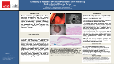
Case Description/Methods: A 75-year-old male with history of COPD and hyperlipidemia presented with longstanding heartburn and dyspepsia. Upper endoscopy (EGD) showed a polypoid, non-circumferential mass on the posterior wall of the stomach. Endoscopic ultrasound (EUS) showed an oval hypoechoic and homogeneous subepithelial lesion originating from the deep mucosa, measuring 15 mm in maximal thickness and 24 mm in diameter with an intact interface between the mass and adjacent structure suggesting a gastrointestinal stromal tumor. After an extended discussion of endoscopic surveillance versus endotherapy, the patient opted for endoscopic resection. Pathology was consistent with gastric duplication cyst (Figure A-D).
Discussion: Gastrointestinal duplication cyst can be defined as a spheroidal structure composed of a muscular layer lined by a mucous membrane. The stomach is the least common site for duplication cysts, accounting for only 2-9% of all cases. The mucosal lining of GDCs is commonly gastric, but ectopic gastrointestinal mucosa including duodenal and pancreatic mucosa has been reported. In our case, the mucosal lining was a mixture of gastric mucosa and ciliated respiratory epithelium, which is an exceedingly rare finding. An accurate diagnosis prior to resection is difficult and particularly so with a polypoid appearance. Differential diagnoses include gastrointestinal stromal tumors, neuroendocrine tumors, and gastric heterotropia. Although there are no established guidelines for managing asymptomatic patients, resection is recommended for symptomatic cases. While most GDCs are benign, it is important to be aware that a small proportion exhibit malignant transformation.

Disclosures:
Kanana Aburayyan, MD1, Raakhi Menon, DO1, Agnes Udoh, MD2, Sreeram Parupudi, MD1. P2804 - Endoscopic Resection of Gastric Duplication Cyst Mimicking Gastrointestinal Stromal Tumor, ACG 2023 Annual Scientific Meeting Abstracts. Vancouver, BC, Canada: American College of Gastroenterology.
1University of Texas Medical Branch, Galveston, TX; 2University of Texas Medical Branch at Galveston, Gavleston, TX
Introduction: Gastric duplication cysts (GDCs) arise during embryonic development and are typically diagnosed in infancy or early childhood. In adults, GDCs are discovered incidentally and are usually asymptomatic. Reported symptoms include nausea, vomiting, dyspepsia and weight loss. We present here an elderly male with non-specific upper abdominal symptoms and endoscopic findings.
Case Description/Methods: A 75-year-old male with history of COPD and hyperlipidemia presented with longstanding heartburn and dyspepsia. Upper endoscopy (EGD) showed a polypoid, non-circumferential mass on the posterior wall of the stomach. Endoscopic ultrasound (EUS) showed an oval hypoechoic and homogeneous subepithelial lesion originating from the deep mucosa, measuring 15 mm in maximal thickness and 24 mm in diameter with an intact interface between the mass and adjacent structure suggesting a gastrointestinal stromal tumor. After an extended discussion of endoscopic surveillance versus endotherapy, the patient opted for endoscopic resection. Pathology was consistent with gastric duplication cyst (Figure A-D).
Discussion: Gastrointestinal duplication cyst can be defined as a spheroidal structure composed of a muscular layer lined by a mucous membrane. The stomach is the least common site for duplication cysts, accounting for only 2-9% of all cases. The mucosal lining of GDCs is commonly gastric, but ectopic gastrointestinal mucosa including duodenal and pancreatic mucosa has been reported. In our case, the mucosal lining was a mixture of gastric mucosa and ciliated respiratory epithelium, which is an exceedingly rare finding. An accurate diagnosis prior to resection is difficult and particularly so with a polypoid appearance. Differential diagnoses include gastrointestinal stromal tumors, neuroendocrine tumors, and gastric heterotropia. Although there are no established guidelines for managing asymptomatic patients, resection is recommended for symptomatic cases. While most GDCs are benign, it is important to be aware that a small proportion exhibit malignant transformation.

Figure: A: EGD showing a polypoid, non-circumferential mass on the posterior wall of the stomach.
B: EUS showing a hypoechoic and homogeneous subepithelial lesion originating from the deep mucosa.
C: Gross appearance of endoscopically removed gastric lesion.
D: Histological appearance of cyst wall lined by tall columnar ciliated epithelium.
E: Histological appearance of full cross section gastric specimen showing cyst wall and adjacent normal gastric mucosa.
Disclosures:
Kanana Aburayyan indicated no relevant financial relationships.
Raakhi Menon indicated no relevant financial relationships.
Agnes Udoh indicated no relevant financial relationships.
Sreeram Parupudi indicated no relevant financial relationships.
Kanana Aburayyan, MD1, Raakhi Menon, DO1, Agnes Udoh, MD2, Sreeram Parupudi, MD1. P2804 - Endoscopic Resection of Gastric Duplication Cyst Mimicking Gastrointestinal Stromal Tumor, ACG 2023 Annual Scientific Meeting Abstracts. Vancouver, BC, Canada: American College of Gastroenterology.
