Monday Poster Session
Category: Stomach
P2808 - Signet Ring Cell Changes in Gastritis - Recognizing a Rare Diagnostic Pitfall
Monday, October 23, 2023
10:30 AM - 4:15 PM PT
Location: Exhibit Hall

Has Audio
- KB
Kuntal Bhowmick, MD
The Warren Alpert Medical School of Brown University
Providence, RI
Presenting Author(s)
Kuntal Bhowmick, MD1, Yousef Elfanagely, MD1, Andrew Krane, MD1, Pranith Perera, MD2
1Warren Alpert Medical School of Brown University, Providence, RI; 2Brown University, Providence, RI
Introduction: Signet ring cells on histology are most often associated with malignant adenocarcinoma. However, signet ring cell changes (SRCC) represent a benign, reactive process, and can be mistaken for malignancy in biopsy specimens. Cases of SRCC are sporadically reported in the literature, and most often documented with pseudomembranous colitis. We present a case of ulcerative gastritis with pathology initially concerning for adenocarcinoma, but was later determined to be SRCC.
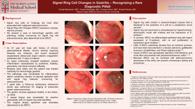
Case Description/Methods: An 81 year old male with a history of chronic gastrointestinal bleeds, chronic anemia requiring biweekly iron infusions, and recent duodenal angioectasia status-post argon plasma coagulation presented to the emergency department with 11 days of melena. An esophagogastroduodenoscopy would reveal scattered, severe inflammation characterized by erythema, friability, and linear erosions found in the cardia, gastric fundus, greater and lesser curvatures of the stomach, incisura, gastric antrum, and pylorus. He was treated medically with a proton pump inhibitor. The pathology from his stomach biopsies returned with the majority of the inflammatory debris containing clusters of atypical epithelial cells with signet ring features, highly suspicious for adenocarcinoma. The patient was scheduled for a follow-up endoscopic ultrasound (EUS) 6 days later for work-up of presumed gastric adenocarcinoma, but his gastric mucosa appeared normal. His EUS was also normal. A biopsy of the gastric antrum was remarkable for reactive gastropathy with no signet ring cells identified. The original biopsy specimen was ultimately determined to be SRCC.
Discussion: Signet ring cells contain a crescent-shaped nucleus that is displaced to the periphery of a cell by a cytoplasmic mucin vacuole. Signet ring cell carcinoma is characterized by hyperchromatic, pleomorphic nuclei with mitoses and low expression of E-cadherin. In contrast, SRCC are differentiated epithelial cells with higher expression of E-cadherin, with no cell proliferation or suppressor gene mutation. These cells potentially develop from an ischemic process, and have been documented in various cases, including a tubular adenoma, gallbladder with cholelithiasis, and Peutz-Jeghers polyp. Therefore, because SRCC can be confused with adenocarcinoma, awareness of the entity can prevent unnecessary testing and procedures.

Disclosures:
Kuntal Bhowmick, MD1, Yousef Elfanagely, MD1, Andrew Krane, MD1, Pranith Perera, MD2. P2808 - Signet Ring Cell Changes in Gastritis - Recognizing a Rare Diagnostic Pitfall, ACG 2023 Annual Scientific Meeting Abstracts. Vancouver, BC, Canada: American College of Gastroenterology.
1Warren Alpert Medical School of Brown University, Providence, RI; 2Brown University, Providence, RI
Introduction: Signet ring cells on histology are most often associated with malignant adenocarcinoma. However, signet ring cell changes (SRCC) represent a benign, reactive process, and can be mistaken for malignancy in biopsy specimens. Cases of SRCC are sporadically reported in the literature, and most often documented with pseudomembranous colitis. We present a case of ulcerative gastritis with pathology initially concerning for adenocarcinoma, but was later determined to be SRCC.
Case Description/Methods: An 81 year old male with a history of chronic gastrointestinal bleeds, chronic anemia requiring biweekly iron infusions, and recent duodenal angioectasia status-post argon plasma coagulation presented to the emergency department with 11 days of melena. An esophagogastroduodenoscopy would reveal scattered, severe inflammation characterized by erythema, friability, and linear erosions found in the cardia, gastric fundus, greater and lesser curvatures of the stomach, incisura, gastric antrum, and pylorus. He was treated medically with a proton pump inhibitor. The pathology from his stomach biopsies returned with the majority of the inflammatory debris containing clusters of atypical epithelial cells with signet ring features, highly suspicious for adenocarcinoma. The patient was scheduled for a follow-up endoscopic ultrasound (EUS) 6 days later for work-up of presumed gastric adenocarcinoma, but his gastric mucosa appeared normal. His EUS was also normal. A biopsy of the gastric antrum was remarkable for reactive gastropathy with no signet ring cells identified. The original biopsy specimen was ultimately determined to be SRCC.
Discussion: Signet ring cells contain a crescent-shaped nucleus that is displaced to the periphery of a cell by a cytoplasmic mucin vacuole. Signet ring cell carcinoma is characterized by hyperchromatic, pleomorphic nuclei with mitoses and low expression of E-cadherin. In contrast, SRCC are differentiated epithelial cells with higher expression of E-cadherin, with no cell proliferation or suppressor gene mutation. These cells potentially develop from an ischemic process, and have been documented in various cases, including a tubular adenoma, gallbladder with cholelithiasis, and Peutz-Jeghers polyp. Therefore, because SRCC can be confused with adenocarcinoma, awareness of the entity can prevent unnecessary testing and procedures.

Figure: A. Endoscopy depicting gastritis of the antrum, which would demonstrate signet ring cell changes on biopsy. B. Follow-up endoscopy with complete resolution of gastritis after 6 days.
Disclosures:
Kuntal Bhowmick indicated no relevant financial relationships.
Yousef Elfanagely indicated no relevant financial relationships.
Andrew Krane indicated no relevant financial relationships.
Pranith Perera indicated no relevant financial relationships.
Kuntal Bhowmick, MD1, Yousef Elfanagely, MD1, Andrew Krane, MD1, Pranith Perera, MD2. P2808 - Signet Ring Cell Changes in Gastritis - Recognizing a Rare Diagnostic Pitfall, ACG 2023 Annual Scientific Meeting Abstracts. Vancouver, BC, Canada: American College of Gastroenterology.
