Monday Poster Session
Category: Stomach
P2819 - Rare Case of Gastric Non-Hodgkin Lymphoma in a Helicobacter pylori-Infected Patient
Monday, October 23, 2023
10:30 AM - 4:15 PM PT
Location: Exhibit Hall

Has Audio

Maram Alenzi, MD
St. Elizabeth's Medical Center
Boston, MA
Presenting Author(s)
Maram Alenzi, MD1, Iyiad AlAbdul-Razzak, MD1, Darren Evanchuk, MD1, Svetlana Kondratiev, MD1, Syed K. Mahmood, MD, MPH2, Manish Tandon, MD1
1St. Elizabeth's Medical Center, Boston, MA; 2St. Elizabeth's Medical Center, Brighton, MA
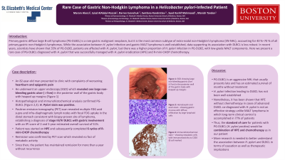
Introduction: Primary gastric diffuse large B-cell lymphoma (PG-DLBCL) is a rare gastric malignant neoplasm, but it is the most common subtype of extra-nodal non-Hodgkin’s lymphoma (EN-NHL), accounting for 40 %–70 % of all primary gastric non-Hodgkin’s lymphomas. While the association between H. pylori infection and gastric MALT lymphoma is well-established, data supporting its association with DLBCL is less robust. In recent years, scientists have shown that 35% of PG-DLBCL patients are affected with H. pylori, but there was a higher proportion of H. pylori infection in PG-DLBCL with low-grade MALT components. Here we present a rare case of PG-DLBCL diagnosed with H. pylori that was successfully managed with H. pylori eradication (HPE) and R-mini-CHOP chemotherapy.
Case Description/Methods: An 82-year-old man presented to clinic with complaints of worsening epigastric pain and heartburn. He underwent an upper endoscopy (EGD) which revealed one large non-bleeding gastric ulcer ( >7cm) in the posterior wall of the gastric body with heaped up margins. Histopathological and immunohistochemical analysis confirmed PG-DLBCL (Figure). H. Pylori stain was positive. Positron emission tomography (PET) scan revealed multiple FDG avid supra and infra-diaphragmatic lymph nodes with focal FDG uptake in the distal stomach consistent with biopsy-proven site of lymphoma, establishing a diagnosis of stage III/IV DLBCL with gastric involvement with an IPI score of 2 and 5-year estimated overall survival of 51%. He was started on HPE and subsequently completed 6 cycles of R-mini-CHOP chemotherapy. Remission was confirmed by PET scan which revealed no foci of metabolic activity.
Discussion: PG-DLBCL is an aggressive NHL that usually presents late and has an estimated survival of months without treatment. H. pylori infection leading to DLBCL without MALT lymphoma features has not been well established. Nonetheless, it has been shown that HPE without chemotherapy in cases of DLBCL co-diagnosed with H. pylori is not an effective strategy unlike MALT lymphoma in which long-term clinical control is accomplished in 77% of patients. Thus, the standard of care for patients with PG-DLBCL (H. pylori positive) would be combination of HPE and chemotherapy as in our patient. More research is needed to better understand the association between H. pylori and DLBCL in terms of causation as well as therapeutic implications.

Disclosures:
Maram Alenzi, MD1, Iyiad AlAbdul-Razzak, MD1, Darren Evanchuk, MD1, Svetlana Kondratiev, MD1, Syed K. Mahmood, MD, MPH2, Manish Tandon, MD1. P2819 - Rare Case of Gastric Non-Hodgkin Lymphoma in a Helicobacter pylori-Infected Patient, ACG 2023 Annual Scientific Meeting Abstracts. Vancouver, BC, Canada: American College of Gastroenterology.
1St. Elizabeth's Medical Center, Boston, MA; 2St. Elizabeth's Medical Center, Brighton, MA
Introduction: Primary gastric diffuse large B-cell lymphoma (PG-DLBCL) is a rare gastric malignant neoplasm, but it is the most common subtype of extra-nodal non-Hodgkin’s lymphoma (EN-NHL), accounting for 40 %–70 % of all primary gastric non-Hodgkin’s lymphomas. While the association between H. pylori infection and gastric MALT lymphoma is well-established, data supporting its association with DLBCL is less robust. In recent years, scientists have shown that 35% of PG-DLBCL patients are affected with H. pylori, but there was a higher proportion of H. pylori infection in PG-DLBCL with low-grade MALT components. Here we present a rare case of PG-DLBCL diagnosed with H. pylori that was successfully managed with H. pylori eradication (HPE) and R-mini-CHOP chemotherapy.
Case Description/Methods: An 82-year-old man presented to clinic with complaints of worsening epigastric pain and heartburn. He underwent an upper endoscopy (EGD) which revealed one large non-bleeding gastric ulcer ( >7cm) in the posterior wall of the gastric body with heaped up margins. Histopathological and immunohistochemical analysis confirmed PG-DLBCL (Figure). H. Pylori stain was positive. Positron emission tomography (PET) scan revealed multiple FDG avid supra and infra-diaphragmatic lymph nodes with focal FDG uptake in the distal stomach consistent with biopsy-proven site of lymphoma, establishing a diagnosis of stage III/IV DLBCL with gastric involvement with an IPI score of 2 and 5-year estimated overall survival of 51%. He was started on HPE and subsequently completed 6 cycles of R-mini-CHOP chemotherapy. Remission was confirmed by PET scan which revealed no foci of metabolic activity.
Discussion: PG-DLBCL is an aggressive NHL that usually presents late and has an estimated survival of months without treatment. H. pylori infection leading to DLBCL without MALT lymphoma features has not been well established. Nonetheless, it has been shown that HPE without chemotherapy in cases of DLBCL co-diagnosed with H. pylori is not an effective strategy unlike MALT lymphoma in which long-term clinical control is accomplished in 77% of patients. Thus, the standard of care for patients with PG-DLBCL (H. pylori positive) would be combination of HPE and chemotherapy as in our patient. More research is needed to better understand the association between H. pylori and DLBCL in terms of causation as well as therapeutic implications.

Figure: Figure 1: (A) EGD showing large non-bleeding gastric ulcer (>7cm) in the posterior wall of the gastric body with heaped up margins. (B) Hematoxylin and eosin stain - showing gastric mucosa with diffuse infiltration by intermediate to large lymphoid cells. (C) Immunohistochemical stain – showing neoplastic cells with immunoreactivity to CD20 (B cell marker).
Disclosures:
Maram Alenzi indicated no relevant financial relationships.
Iyiad AlAbdul-Razzak indicated no relevant financial relationships.
Darren Evanchuk indicated no relevant financial relationships.
Svetlana Kondratiev indicated no relevant financial relationships.
Syed Mahmood indicated no relevant financial relationships.
Manish Tandon indicated no relevant financial relationships.
Maram Alenzi, MD1, Iyiad AlAbdul-Razzak, MD1, Darren Evanchuk, MD1, Svetlana Kondratiev, MD1, Syed K. Mahmood, MD, MPH2, Manish Tandon, MD1. P2819 - Rare Case of Gastric Non-Hodgkin Lymphoma in a Helicobacter pylori-Infected Patient, ACG 2023 Annual Scientific Meeting Abstracts. Vancouver, BC, Canada: American College of Gastroenterology.
