Monday Poster Session
Category: Stomach
P2821 - Cerebellar Involvement as a Rare Manifestation of Gastric Cancer Metastasis
Monday, October 23, 2023
10:30 AM - 4:15 PM PT
Location: Exhibit Hall

Has Audio

Farhan Azad, DO
University at Buffalo
Buffalo, New York
Presenting Author(s)
Farhan Azad, DO, Alexander M. Carlson, DO, Clive J. Miranda, DO, MS, Ross R. Moyer, DO, Prutha Patel, DO, Muddasir Ayaz, MD
University at Buffalo, Buffalo, NY
Introduction: Gastric adenocarcinoma is the fourth most frequent cause of cancer-associated death worldwide. Metastasis is seen in 1/3 of the cases. Central nervous system (CNS) metastases are rare and occur in 0.16–0.69% of patients with gastric or gastroesophageal junction cancer (GEC). We report a case of a patient with newly diagnosed GEC, presenting with symptomatic isolated brain metastases (BM), treated successfully with stereotactic radiosurgery (SRS).
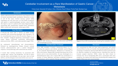
Case Description/Methods: A 69-year-old male with a history of GERD on omeprazole initially presented with progressively worsening dysphagia. EGD revealed a 35 cm circumferential mass at the GE junction (Figure 1). A EUS revealed malignant-appearing paratracheal lymph nodes. Biopsy confirmed moderately differentiated adenocarcinoma, HER2+, and endoscopically staged T3N1. He underwent chemotherapy and chemoradiation, followed by esophagectomy with a biopsy showing residual invasive adenocarcinoma and a partial therapeutic response. Adjuvant immunotherapy with nivolumab was started. One month after starting nivolumab and eight months after his initial diagnosis, he presented with progressively worsening unstable gait, nausea, and headaches. The patient was afebrile and hemodynamically stable. Laboratory values were unrevealing of anemia or increased WBCs, with normal kidney and liver functions. The patient was alert and oriented, with no focal deficits and the Glasgow Coma Scale (GCS) score was 15. Brain MRI revealed 2 lesions, measuring about 2.5 cm, involving the superior and inferior cerebellar vermis (Figure 2). He received dexamethasone, followed by 3 fractions of SRS to the lesions. Repeat MRI revealed decreased size of the lesions, with no new abnormalities. The patient was discharged on a dexamethasone taper, with plans for a repeat MRI in 2 months. CT chest, abdomen, and pelvis revealed no evidence of metastases, and no further systemic therapy was considered.
Discussion: GEC complicated by BM can have rapid progression and overall survival (OS) of as low as 3 months. No standard guidelines exist for screening or treatment in these cases. Gamma-knife SRS has shown promise to be an effective modality for localized lesions, obviating the need for invasive surgical resection. HER2+ status, as seen here, has been shown to increase the risk of developing BM and is associated with poor prognosis. Our patient will need close monitoring with imaging, and regular follow-ups were recommended.

Disclosures:
Farhan Azad, DO, Alexander M. Carlson, DO, Clive J. Miranda, DO, MS, Ross R. Moyer, DO, Prutha Patel, DO, Muddasir Ayaz, MD. P2821 - Cerebellar Involvement as a Rare Manifestation of Gastric Cancer Metastasis, ACG 2023 Annual Scientific Meeting Abstracts. Vancouver, BC, Canada: American College of Gastroenterology.
University at Buffalo, Buffalo, NY
Introduction: Gastric adenocarcinoma is the fourth most frequent cause of cancer-associated death worldwide. Metastasis is seen in 1/3 of the cases. Central nervous system (CNS) metastases are rare and occur in 0.16–0.69% of patients with gastric or gastroesophageal junction cancer (GEC). We report a case of a patient with newly diagnosed GEC, presenting with symptomatic isolated brain metastases (BM), treated successfully with stereotactic radiosurgery (SRS).
Case Description/Methods: A 69-year-old male with a history of GERD on omeprazole initially presented with progressively worsening dysphagia. EGD revealed a 35 cm circumferential mass at the GE junction (Figure 1). A EUS revealed malignant-appearing paratracheal lymph nodes. Biopsy confirmed moderately differentiated adenocarcinoma, HER2+, and endoscopically staged T3N1. He underwent chemotherapy and chemoradiation, followed by esophagectomy with a biopsy showing residual invasive adenocarcinoma and a partial therapeutic response. Adjuvant immunotherapy with nivolumab was started. One month after starting nivolumab and eight months after his initial diagnosis, he presented with progressively worsening unstable gait, nausea, and headaches. The patient was afebrile and hemodynamically stable. Laboratory values were unrevealing of anemia or increased WBCs, with normal kidney and liver functions. The patient was alert and oriented, with no focal deficits and the Glasgow Coma Scale (GCS) score was 15. Brain MRI revealed 2 lesions, measuring about 2.5 cm, involving the superior and inferior cerebellar vermis (Figure 2). He received dexamethasone, followed by 3 fractions of SRS to the lesions. Repeat MRI revealed decreased size of the lesions, with no new abnormalities. The patient was discharged on a dexamethasone taper, with plans for a repeat MRI in 2 months. CT chest, abdomen, and pelvis revealed no evidence of metastases, and no further systemic therapy was considered.
Discussion: GEC complicated by BM can have rapid progression and overall survival (OS) of as low as 3 months. No standard guidelines exist for screening or treatment in these cases. Gamma-knife SRS has shown promise to be an effective modality for localized lesions, obviating the need for invasive surgical resection. HER2+ status, as seen here, has been shown to increase the risk of developing BM and is associated with poor prognosis. Our patient will need close monitoring with imaging, and regular follow-ups were recommended.

Figure: Figure 1: EGD showing a 35 cm circumferential mass at the gastroesophageal junction
Figure 2: Brain MRI showing lesion involving the cerebellar vermis
Figure 2: Brain MRI showing lesion involving the cerebellar vermis
Disclosures:
Farhan Azad indicated no relevant financial relationships.
Alexander Carlson indicated no relevant financial relationships.
Clive Miranda indicated no relevant financial relationships.
Ross Moyer indicated no relevant financial relationships.
Prutha Patel indicated no relevant financial relationships.
Muddasir Ayaz indicated no relevant financial relationships.
Farhan Azad, DO, Alexander M. Carlson, DO, Clive J. Miranda, DO, MS, Ross R. Moyer, DO, Prutha Patel, DO, Muddasir Ayaz, MD. P2821 - Cerebellar Involvement as a Rare Manifestation of Gastric Cancer Metastasis, ACG 2023 Annual Scientific Meeting Abstracts. Vancouver, BC, Canada: American College of Gastroenterology.
