Monday Poster Session
Category: Stomach
P2831 - Isolated Gastric Metastasis of Renal Cell Carcinoma 11 Years After Initial Diagnosis Treated With Endoscopic Submucosal Dissection
Monday, October 23, 2023
10:30 AM - 4:15 PM PT
Location: Exhibit Hall

Has Audio
- MO
Mark Obri, MD
Henry Ford Hospital
Detroit, Michigan
Presenting Author(s)
Mark Obri, MD, Muhammad Zarrar Khan, MD, Ammad Chaudhary, MD, Cyrus Piraka, MD, Tobias Zuchelli, MD
Henry Ford Hospital, Detroit, MI
Introduction: Renal Cell Carcinoma (RCC) is the most common form of malignant renal disease with high incidence throughout the world. The incidence of gastric metastasis has been reported to be less than 5%, with isolated gastric metastasis less likely. Prior literature reports the average diagnoses of gastric metastasis 7 years after initial presentation. Here, we present a case of isolated gastric metastasis of RCC that presented 11 years after diagnosis of RCC.
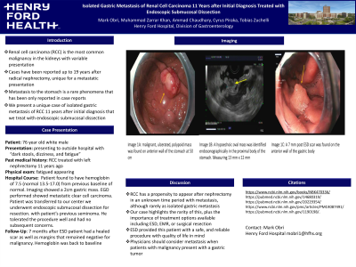
Case Description/Methods: A 76 y/o white male with past medical history of renal cell carcinoma treated with left nephrectomy 11 years ago presented to an outside hospital with dark stools, dizziness and fatigue. He was found to have a low hemoglobin and subsequent Computed Tomography (CT) with IV contrast was inconclusive. He underwent an upper endoscopy and endoscopic ultrasound, which showed a 2 cm gastric mass in the proximal gastric body along the greater curvature (Image 1). Biopsy of the mass showed metastatic clear cell renal carcinoma, which is what the patient had been diagnosed with 11 years prior. A positron emission tomography (PET) scan showed uptake in the stomach but no evidence of distant metastasis.
The patient was referred to our center for endoscopic submucosal dissection (ESD) for resection of the mass. Hybrid ESD was used to remove the lesion en bloc. Resection margins were negative. Follow-up endoscopy 7 months later showed the stomach and margins remained negative for malignancy and the patient was asymptomatic.
Discussion: We present a case with a rare presentation of isolated gastric metastasis from RCC 11 years after initial diagnosis. This is later than the average of 7 years reported in the literature and is significant due to its isolated gastric nature. We also offer a non-surgical approach with ESD which the patient tolerated well with clean margins almost 1 year later.

Disclosures:
Mark Obri, MD, Muhammad Zarrar Khan, MD, Ammad Chaudhary, MD, Cyrus Piraka, MD, Tobias Zuchelli, MD. P2831 - Isolated Gastric Metastasis of Renal Cell Carcinoma 11 Years After Initial Diagnosis Treated With Endoscopic Submucosal Dissection, ACG 2023 Annual Scientific Meeting Abstracts. Vancouver, BC, Canada: American College of Gastroenterology.
Henry Ford Hospital, Detroit, MI
Introduction: Renal Cell Carcinoma (RCC) is the most common form of malignant renal disease with high incidence throughout the world. The incidence of gastric metastasis has been reported to be less than 5%, with isolated gastric metastasis less likely. Prior literature reports the average diagnoses of gastric metastasis 7 years after initial presentation. Here, we present a case of isolated gastric metastasis of RCC that presented 11 years after diagnosis of RCC.
Case Description/Methods: A 76 y/o white male with past medical history of renal cell carcinoma treated with left nephrectomy 11 years ago presented to an outside hospital with dark stools, dizziness and fatigue. He was found to have a low hemoglobin and subsequent Computed Tomography (CT) with IV contrast was inconclusive. He underwent an upper endoscopy and endoscopic ultrasound, which showed a 2 cm gastric mass in the proximal gastric body along the greater curvature (Image 1). Biopsy of the mass showed metastatic clear cell renal carcinoma, which is what the patient had been diagnosed with 11 years prior. A positron emission tomography (PET) scan showed uptake in the stomach but no evidence of distant metastasis.
The patient was referred to our center for endoscopic submucosal dissection (ESD) for resection of the mass. Hybrid ESD was used to remove the lesion en bloc. Resection margins were negative. Follow-up endoscopy 7 months later showed the stomach and margins remained negative for malignancy and the patient was asymptomatic.
Discussion: We present a case with a rare presentation of isolated gastric metastasis from RCC 11 years after initial diagnosis. This is later than the average of 7 years reported in the literature and is significant due to its isolated gastric nature. We also offer a non-surgical approach with ESD which the patient tolerated well with clean margins almost 1 year later.

Figure: Image 1A: Malignant, ulcerated polypoid mass was found on anterior wall of the stomach at 50cm
Image 1B: A hypoechoic oval mass was identified endosonographically in the proximal body of the stomach. Measuring 13mm x 12mm
Image 1C: A 7mm post ESD scar was found on the anterior wall of the gastric body
Image 1B: A hypoechoic oval mass was identified endosonographically in the proximal body of the stomach. Measuring 13mm x 12mm
Image 1C: A 7mm post ESD scar was found on the anterior wall of the gastric body
Disclosures:
Mark Obri indicated no relevant financial relationships.
Muhammad Zarrar Khan indicated no relevant financial relationships.
Ammad Chaudhary indicated no relevant financial relationships.
Cyrus Piraka: Boston Scientific – Consultant.
Tobias Zuchelli: Boston Scientific – Consultant.
Mark Obri, MD, Muhammad Zarrar Khan, MD, Ammad Chaudhary, MD, Cyrus Piraka, MD, Tobias Zuchelli, MD. P2831 - Isolated Gastric Metastasis of Renal Cell Carcinoma 11 Years After Initial Diagnosis Treated With Endoscopic Submucosal Dissection, ACG 2023 Annual Scientific Meeting Abstracts. Vancouver, BC, Canada: American College of Gastroenterology.
