Monday Poster Session
Category: Stomach
P2832 - A Collagenous Conundrum
Monday, October 23, 2023
10:30 AM - 4:15 PM PT
Location: Exhibit Hall

Has Audio
- JA
Jamil O. Alexis, MD
Bridgeport Hospital/Yale New Haven Health
Bridgeport, CT
Presenting Author(s)
Jamil O. Alexis, MD1, Prince Addo. Ameyaw, MD1, Joanna Gibson, MD, PhD2, Benjamin Barbash, MD3
1Bridgeport Hospital/Yale New Haven Health, Bridgeport, CT; 2Yale University School of Medicine, New Haven, CT; 3Bridgeport Hospital, Trumbull, CT
Introduction: Collagenous gastritis (CG) is an uncommon pathologic condition characterized by the presence of distinct subepithelial collagenous band within the gastric mucosa. The clinical presentation of CG can vary widely ranging from nonspecific gastrointestinal symptoms to severe anemia and nutritional deficiencies. We present a patient with CG to highlight management strategies and the subsequent clinical outcome. Through this patient case, we aim to contribute to existing knowledge surrounding CG and emphasize the importance of considering this rare condition when evaluating patient with unexplained anemia without typical gastrointestinal manifestations associated with this disease entity.
Case Description/Methods: A 21 year old male presented for outpatient evaluation of microcytic anemia. He denied any abdominal symptoms, weight loss, or overt GI bleeding. Physical examination revealed stable vital signs, normal gastrointestinal and cardiovascular findings. Labs revealed Hgb 10.3, MCV 66.1, Ferritin 3, Total Iron 19, TIBC 489, Calprotectin 6, CRP 0.7, negative workup for hemoglobinopathies and celiac disease. EGD showed diffuse nodular mucosa of the gastric body, patchy erythematous prepyloric mucosa. Biopsies of the gastric antrum/body showed increased eosinophils >50/hpf, thickened subepithelial collagen deposition and negative H. pylori immunostain. Duodenal biopsies were normal. Colonoscopy and subsequent MR enterography were unremarkable. Patient was diagnosed with collagenous gastritis and managed with pantoprazole 40mg daily, iron supplementation, open capsule budesonide 3mg TID for 4 months followed by a 2 month taper. Hemoglobin and iron studies normalized 2 months into treatment with budesonide.
Discussion: Collagenous gastritis has been characterized as two different phenotypes, adult and pediatric. The symptomology of the pediatric type include abdominal pain and GI bleeding, while the adult type can present with chronic diarrhea, weight loss, and sequelae from associated nutritional deficiencies. This patient atypically had an asymptomatic presentation, with iron deficiency anemia being the only abnormal lab finding. Endoscopy revealed expected typical findings of nodular gastric mucosa and mucosal erythema. No standard therapy exist due to its rarity. Of the known treatment options, steroids, in particular open capsule budesonide, coupled with iron supplementation, helped our patient achieve quick and complete clinical resolution of his anemia.

Disclosures:
Jamil O. Alexis, MD1, Prince Addo. Ameyaw, MD1, Joanna Gibson, MD, PhD2, Benjamin Barbash, MD3. P2832 - A Collagenous Conundrum, ACG 2023 Annual Scientific Meeting Abstracts. Vancouver, BC, Canada: American College of Gastroenterology.
1Bridgeport Hospital/Yale New Haven Health, Bridgeport, CT; 2Yale University School of Medicine, New Haven, CT; 3Bridgeport Hospital, Trumbull, CT
Introduction: Collagenous gastritis (CG) is an uncommon pathologic condition characterized by the presence of distinct subepithelial collagenous band within the gastric mucosa. The clinical presentation of CG can vary widely ranging from nonspecific gastrointestinal symptoms to severe anemia and nutritional deficiencies. We present a patient with CG to highlight management strategies and the subsequent clinical outcome. Through this patient case, we aim to contribute to existing knowledge surrounding CG and emphasize the importance of considering this rare condition when evaluating patient with unexplained anemia without typical gastrointestinal manifestations associated with this disease entity.
Case Description/Methods: A 21 year old male presented for outpatient evaluation of microcytic anemia. He denied any abdominal symptoms, weight loss, or overt GI bleeding. Physical examination revealed stable vital signs, normal gastrointestinal and cardiovascular findings. Labs revealed Hgb 10.3, MCV 66.1, Ferritin 3, Total Iron 19, TIBC 489, Calprotectin 6, CRP 0.7, negative workup for hemoglobinopathies and celiac disease. EGD showed diffuse nodular mucosa of the gastric body, patchy erythematous prepyloric mucosa. Biopsies of the gastric antrum/body showed increased eosinophils >50/hpf, thickened subepithelial collagen deposition and negative H. pylori immunostain. Duodenal biopsies were normal. Colonoscopy and subsequent MR enterography were unremarkable. Patient was diagnosed with collagenous gastritis and managed with pantoprazole 40mg daily, iron supplementation, open capsule budesonide 3mg TID for 4 months followed by a 2 month taper. Hemoglobin and iron studies normalized 2 months into treatment with budesonide.
Discussion: Collagenous gastritis has been characterized as two different phenotypes, adult and pediatric. The symptomology of the pediatric type include abdominal pain and GI bleeding, while the adult type can present with chronic diarrhea, weight loss, and sequelae from associated nutritional deficiencies. This patient atypically had an asymptomatic presentation, with iron deficiency anemia being the only abnormal lab finding. Endoscopy revealed expected typical findings of nodular gastric mucosa and mucosal erythema. No standard therapy exist due to its rarity. Of the known treatment options, steroids, in particular open capsule budesonide, coupled with iron supplementation, helped our patient achieve quick and complete clinical resolution of his anemia.

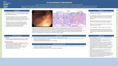
Figure: Endoscopic view of the nodular gastric mucosa in Image A. Histologic examination of stomach (body) biopsy, with hematoxylin and eosin (H&E) stain in Image B, trichrome stain depicted in Image C (200x magnification). The H&E stain (Image B) demonstrates expansion of the lamina propria by chronic inflammatory cells (stars) and increased eosinophils (arrow head) between the onxyntic glands (G). At the mucosal surface, the foveolar epithelium (FE) is degenerated, with loss of intracellular mucin vacuoles and detachment. The subepithelial collagen layer is markedly expanded and hyalinized (brackets), with embedded inflammatory cells. The trichrome stain (Image C) highlights the thickened collagen layer (blue). There is no Helicobacter pylori, intestinal metaplasia or atrophy identified. The overall features are consistent with collagenous gastritis.
Disclosures:
Jamil Alexis indicated no relevant financial relationships.
Prince Ameyaw indicated no relevant financial relationships.
Joanna Gibson indicated no relevant financial relationships.
Benjamin Barbash indicated no relevant financial relationships.
Jamil O. Alexis, MD1, Prince Addo. Ameyaw, MD1, Joanna Gibson, MD, PhD2, Benjamin Barbash, MD3. P2832 - A Collagenous Conundrum, ACG 2023 Annual Scientific Meeting Abstracts. Vancouver, BC, Canada: American College of Gastroenterology.
