Monday Poster Session
Category: Stomach
P2836 - A Rare Case of Gastric Small Cell Carcinoma Presenting With Severe Hypercalcemia and Liver Metastases
Monday, October 23, 2023
10:30 AM - 4:15 PM PT
Location: Exhibit Hall

Has Audio

Nigil Thaimuriyil, DO
Baptist Hospitals of Southeast Texas
Beaumont, TX
Presenting Author(s)
Ahmed Abdelhakeem, MD1, Syed Jilani, MD1, Nigil Thaimuriyil, DO2, Amr Hamed, MD1, Tahir Naqvi, MD3
1Baptist Hospitals of Southeast Texas, Beaumont, TX; 2Baptist Hospitals of Southeast Texas, Newark, NJ; 3Baptist Physicians Network, Beaumont, TX
Introduction: Gastric small cell carcinomas (GSCC) are very rare tumors characterized by their aggressive nature and poor prognosis. They commonly present with regional lymph nodes involvement and metastases to distant organs. Initial symptoms are usually non-specific, including weight loss, fatigue, and anorexia. However, some GSCC may present with excess hormone secretion, such as parathyroid hormone-related protein (PTHrP), serotonin, and calcitonin. We present a case of metastatic GSCC with initial presentation of severe hypercalcemia.
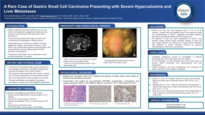
Case Description/Methods: A 73-year-old Caucasian female patient presented with dizziness and fatigue associated with nausea, vomiting, and weight loss. Laboratory evaluation showed severe hypercalcemia (20.2 mg/dL) with low PTH (5 pg/mL )and high PTHrP (16.1 pMOL/L). CT of the abdomen showed a large gastric mass with enlarged regional lymph nodes enlargement and many hepatic lesions. EGD showed a large mass in the gastric body. Biopsy was positive for small cell neuroendocrine carcinomas of primary gastric origin. Immune satins were positive for pan-cytokeratin (AE1/AE3), synaptophysin, chromogranin, and negative staining for CD56, S-100, HMB-45, Melan-A, and CD117, TTF-1. 80% positive for Ki-67. IV fluid resuscitation and Zoledronic acid infusion were initiated. The patient was offered urgent palliative chemotherapy for stage IV disease. However, with the rapid decline in her performance status, she opted for hospice care.
Discussion: GSCCs are rare cancers representing only 0.1-1% of all primary gastric cancers. Limited data about the histopathology of GSCC suggest similarities between pulmonary and gastrointestinal small-cell cancers. Both forms of SCC feature common histological and immunohistochemical findings such as small round blue cells with scant cytoplasm forming sheets and nests, a high proliferative rate with abundant necrosis, and positive stain for synaptophysin, chromogranin, and CD56. There is still no staging system for GSCC, but it is recommended to perform chest imaging to rule out primary pulmonary SCC. To our knowledge, this is one of the rare cases of primary GSCC presenting with hypercalcemia and liver metastases. Due to the paucity of documented cases, no standard of care has been established for GSCCs, but patients have been offered palliative or radical resection followed by adjuvant chemotherapy if able to tolerate it. Further studies and literature are needed to help guide diagnosis, staging and treatment of this rare but deadly form of SCC.

Disclosures:
Ahmed Abdelhakeem, MD1, Syed Jilani, MD1, Nigil Thaimuriyil, DO2, Amr Hamed, MD1, Tahir Naqvi, MD3. P2836 - A Rare Case of Gastric Small Cell Carcinoma Presenting With Severe Hypercalcemia and Liver Metastases, ACG 2023 Annual Scientific Meeting Abstracts. Vancouver, BC, Canada: American College of Gastroenterology.
1Baptist Hospitals of Southeast Texas, Beaumont, TX; 2Baptist Hospitals of Southeast Texas, Newark, NJ; 3Baptist Physicians Network, Beaumont, TX
Introduction: Gastric small cell carcinomas (GSCC) are very rare tumors characterized by their aggressive nature and poor prognosis. They commonly present with regional lymph nodes involvement and metastases to distant organs. Initial symptoms are usually non-specific, including weight loss, fatigue, and anorexia. However, some GSCC may present with excess hormone secretion, such as parathyroid hormone-related protein (PTHrP), serotonin, and calcitonin. We present a case of metastatic GSCC with initial presentation of severe hypercalcemia.
Case Description/Methods: A 73-year-old Caucasian female patient presented with dizziness and fatigue associated with nausea, vomiting, and weight loss. Laboratory evaluation showed severe hypercalcemia (20.2 mg/dL) with low PTH (5 pg/mL )and high PTHrP (16.1 pMOL/L). CT of the abdomen showed a large gastric mass with enlarged regional lymph nodes enlargement and many hepatic lesions. EGD showed a large mass in the gastric body. Biopsy was positive for small cell neuroendocrine carcinomas of primary gastric origin. Immune satins were positive for pan-cytokeratin (AE1/AE3), synaptophysin, chromogranin, and negative staining for CD56, S-100, HMB-45, Melan-A, and CD117, TTF-1. 80% positive for Ki-67. IV fluid resuscitation and Zoledronic acid infusion were initiated. The patient was offered urgent palliative chemotherapy for stage IV disease. However, with the rapid decline in her performance status, she opted for hospice care.
Discussion: GSCCs are rare cancers representing only 0.1-1% of all primary gastric cancers. Limited data about the histopathology of GSCC suggest similarities between pulmonary and gastrointestinal small-cell cancers. Both forms of SCC feature common histological and immunohistochemical findings such as small round blue cells with scant cytoplasm forming sheets and nests, a high proliferative rate with abundant necrosis, and positive stain for synaptophysin, chromogranin, and CD56. There is still no staging system for GSCC, but it is recommended to perform chest imaging to rule out primary pulmonary SCC. To our knowledge, this is one of the rare cases of primary GSCC presenting with hypercalcemia and liver metastases. Due to the paucity of documented cases, no standard of care has been established for GSCCs, but patients have been offered palliative or radical resection followed by adjuvant chemotherapy if able to tolerate it. Further studies and literature are needed to help guide diagnosis, staging and treatment of this rare but deadly form of SCC.

Figure: A) Esophagogastroduodenoscopy showing a large mass in the gastric body
B) H&E staining showing necrotic tumor cells and increased mitosis
C) Ki-67 staining for tumor cells
B) H&E staining showing necrotic tumor cells and increased mitosis
C) Ki-67 staining for tumor cells
Disclosures:
Ahmed Abdelhakeem indicated no relevant financial relationships.
Syed Jilani indicated no relevant financial relationships.
Nigil Thaimuriyil indicated no relevant financial relationships.
Amr Hamed indicated no relevant financial relationships.
Tahir Naqvi indicated no relevant financial relationships.
Ahmed Abdelhakeem, MD1, Syed Jilani, MD1, Nigil Thaimuriyil, DO2, Amr Hamed, MD1, Tahir Naqvi, MD3. P2836 - A Rare Case of Gastric Small Cell Carcinoma Presenting With Severe Hypercalcemia and Liver Metastases, ACG 2023 Annual Scientific Meeting Abstracts. Vancouver, BC, Canada: American College of Gastroenterology.
