Monday Poster Session
Category: GI Bleeding
P2101 - Timing the Second Look Endoscopy to Find the Elusive Dieulafoy Lesion
Monday, October 23, 2023
10:30 AM - 4:15 PM PT
Location: Exhibit Hall

Has Audio

Temesgen Shibre, MD
Capital Health Regional Medical Center
Marshfield, WI
Presenting Author(s)
Temesgen Shibre, MD1, Deep Mehta, MD2, Abdulkerim Mohammed, MD2, Kaushal Majmudar, MD3
1Capital Health Regional Medical Center, Hamilton, NJ; 2Capital Health Regional Medical Center, Trenton, NJ; 3Capital Health Gastroenterology, Bordentown, NJ
Introduction: Dieulafoy’s Lesion (DL) is a rare and sometimes challenging cause of upper gastrointestinal bleeding (UGIB) to identify. Initial esophagogastroduodenoscopy (EGD) is nondiagnostic in 30% of cases which emphasizes the importance of second look endoscopy in successful diagnosis and management of DL. Here, we present a case of a DL causing brisk UGIB in a patient who had upper and lower gastrointestinal endoscopy for dysphagia and weight loss mere hours before developing brisk UGIB from a DL that was not visualized on index endoscopy.
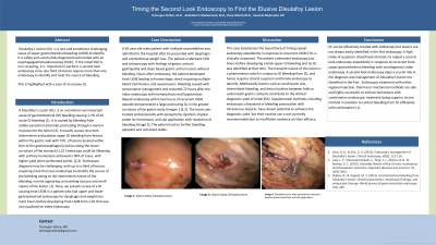
Case Description/Methods: A 65-year-old male patient with multiple comorbidities presented with dysphagia and unintentional weight loss. The patient underwent EGD and colonoscopy with findings of gastric antrum gastropathy and clean based gastric antrum ulcers without bleeding. Hours after endoscopy, the patient developed brisk UGIB leading to hemorrhagic shock requiring multiple blood transfusions and vasopressors. Bleeding ceased with conservative management and restarted 72 hours after the index endoscopy with hematochezia and hypotension. Repeat endoscopy within two hours of recurrent UGIB episode demonstrated a large protruding DL in the greater curvature of the gastric body. The lesion was treated endoscopically with epinephrine injection, bipolar probe for hemostasis, and clip application with resolution of bleeding. The patient had no further bleeding episodes and remained stable.
Discussion: This case emphasizes the importance of timing repeat endoscopy expediently in response to recurrent UGIB if DL is clinically suspected. The patient underwent endoscopy just hours before development of brisk UGIB and no DL was identified at that time. The transient nature of this lesion is a phenomenon which is unique to UGIB from DL, and hence requires clinical suspicion and timely endoscopy to identify. Additionally, factors such as small lesion size, intermittent bleeding, and lesion location between folds or underneath gastric contents contribute to the limited diagnostic yield of initial EGD. Supplemental methods, including endoscopic ultrasound or bleeding provocation with intravenous heparin, have shown potential to enhance the diagnostic yield, but their routine use is not currently recommended due to insufficient evidence on their efficacy.
Second look endoscopy performed expediently in response to recurrent brisk upper gastrointestinal bleeding with nondiagnostic index endoscopy plays a crucial role in the diagnosis and management of Dieulafoy’s lesion.

Disclosures:
Temesgen Shibre, MD1, Deep Mehta, MD2, Abdulkerim Mohammed, MD2, Kaushal Majmudar, MD3. P2101 - Timing the Second Look Endoscopy to Find the Elusive Dieulafoy Lesion, ACG 2023 Annual Scientific Meeting Abstracts. Vancouver, BC, Canada: American College of Gastroenterology.
1Capital Health Regional Medical Center, Hamilton, NJ; 2Capital Health Regional Medical Center, Trenton, NJ; 3Capital Health Gastroenterology, Bordentown, NJ
Introduction: Dieulafoy’s Lesion (DL) is a rare and sometimes challenging cause of upper gastrointestinal bleeding (UGIB) to identify. Initial esophagogastroduodenoscopy (EGD) is nondiagnostic in 30% of cases which emphasizes the importance of second look endoscopy in successful diagnosis and management of DL. Here, we present a case of a DL causing brisk UGIB in a patient who had upper and lower gastrointestinal endoscopy for dysphagia and weight loss mere hours before developing brisk UGIB from a DL that was not visualized on index endoscopy.
Case Description/Methods: A 65-year-old male patient with multiple comorbidities presented with dysphagia and unintentional weight loss. The patient underwent EGD and colonoscopy with findings of gastric antrum gastropathy and clean based gastric antrum ulcers without bleeding. Hours after endoscopy, the patient developed brisk UGIB leading to hemorrhagic shock requiring multiple blood transfusions and vasopressors. Bleeding ceased with conservative management and restarted 72 hours after the index endoscopy with hematochezia and hypotension. Repeat endoscopy within two hours of recurrent UGIB episode demonstrated a large protruding DL in the greater curvature of the gastric body. The lesion was treated endoscopically with epinephrine injection, bipolar probe for hemostasis, and clip application with resolution of bleeding. The patient had no further bleeding episodes and remained stable.
Discussion: This case emphasizes the importance of timing repeat endoscopy expediently in response to recurrent UGIB if DL is clinically suspected. The patient underwent endoscopy just hours before development of brisk UGIB and no DL was identified at that time. The transient nature of this lesion is a phenomenon which is unique to UGIB from DL, and hence requires clinical suspicion and timely endoscopy to identify. Additionally, factors such as small lesion size, intermittent bleeding, and lesion location between folds or underneath gastric contents contribute to the limited diagnostic yield of initial EGD. Supplemental methods, including endoscopic ultrasound or bleeding provocation with intravenous heparin, have shown potential to enhance the diagnostic yield, but their routine use is not currently recommended due to insufficient evidence on their efficacy.
Second look endoscopy performed expediently in response to recurrent brisk upper gastrointestinal bleeding with nondiagnostic index endoscopy plays a crucial role in the diagnosis and management of Dieulafoy’s lesion.

Figure: A & B: Gastric Body: Dieulafoy Lesion
C: Gastric Body Dieulafoy Lesion after epinephrine injection
C: Gastric Body Dieulafoy Lesion after epinephrine injection
Disclosures:
Temesgen Shibre indicated no relevant financial relationships.
Deep Mehta indicated no relevant financial relationships.
Abdulkerim Mohammed indicated no relevant financial relationships.
Kaushal Majmudar indicated no relevant financial relationships.
Temesgen Shibre, MD1, Deep Mehta, MD2, Abdulkerim Mohammed, MD2, Kaushal Majmudar, MD3. P2101 - Timing the Second Look Endoscopy to Find the Elusive Dieulafoy Lesion, ACG 2023 Annual Scientific Meeting Abstracts. Vancouver, BC, Canada: American College of Gastroenterology.
