Monday Poster Session
Category: GI Bleeding
P2103 - A B-Wildering Case of Gastrointestinal Bleeding
Monday, October 23, 2023
10:30 AM - 4:15 PM PT
Location: Exhibit Hall

Has Audio
- AV
Adam Vinall, MD
University of Texas Health Science Center at San Antonio
San Antonio, TX
Presenting Author(s)
Adam Vinall, MD1, Priyadarshini Loganathan, MD2, Mahesh Gajendran, MD, MPH1, Sylvia Keiser, DO1, Alia Nazarullah, MD1, Hari Sayana, MBBS, MD1
1University of Texas Health Science Center at San Antonio, San Antonio, TX; 2University Health System, San Antonio, TX
Introduction: Primary malignant tumors of the small intestine account for less than 2% of all gastrointestinal (GI) neoplasms and typically present with nonspecific symptoms, such as abdominal pain, nausea, and weight loss. We present an unusual case of GI bleeding caused by primary small bowel diffuse large B-cell lymphoma (DLBCL).
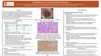
Case Description/Methods: A 63-year-old male with a history of coronary artery disease, on aspirin and clopidogrel, presented to the advanced endoscopy clinic for evaluation of GI bleeding with associated anemia. The patient complained of worsening fatigue, melena, and hematochezia. Chart review showed repeated emergency department visits for symptomatic anemia requiring blood transfusions and a positive Cologuard one year prior. Upper and lower endoscopies, tagged RBC scan, and small bowel follow through tests all were unable to find the bleeding source. Laboratory results were significant for a hemoglobin of 8.5, mean corpuscular volume of 81.5, and normal white blood cell count. The patient underwent retrograde single balloon enteroscopy, which revealed a single, 10-15cm, friable, ulcerated lesion in the distal ileum, 30cm from the ileocecal valve (Figure 1a). Biopsies demonstrated diffuse large B-cell lymphoma, germinal center B-cell phenotype (Figure 1b). Staining was MYC positive, Bcl-2 and Bcl-6 negative. The patient currently follows with hematology for treatment.
Discussion: Primary malignant tumors of the small intestine (SI) are very rare, with less than 2% of GI malignancy constituting small bowel neoplasms. The ileum is the most common site (60%-65%) for SI lymphoma, followed by the jejunum (20%-25%), duodenum (6%-8%), and other sites (8%-9%). SI lymphomas constitute 20% to 30% of all GI lymphomas and 15% to 20% of all SI neoplasms. Common risk factors for SI lymphoma include H. pylori infection, viral infections, inflammatory bowel disease, and immunosuppression.
This cancer typically presents with non-specific symptoms, though rarely presents with acute obstructive symptoms or perforation. The diagnosis of primary GI lymphoma requires the absence of systemic lymphadenopathy and normal white blood cell count on presentation. Currently, capsule endoscopy and balloon enteroscopy are the mainstay of diagnosis, though imaging like PET-CT is becoming increasingly utilized. This case demonstrates the need for high index of clinical suspicion of SI lymphoma in cases of occult GI bleeding and the utility of enteroscopy in the diagnosis of this disease.

Disclosures:
Adam Vinall, MD1, Priyadarshini Loganathan, MD2, Mahesh Gajendran, MD, MPH1, Sylvia Keiser, DO1, Alia Nazarullah, MD1, Hari Sayana, MBBS, MD1. P2103 - A B-Wildering Case of Gastrointestinal Bleeding, ACG 2023 Annual Scientific Meeting Abstracts. Vancouver, BC, Canada: American College of Gastroenterology.
1University of Texas Health Science Center at San Antonio, San Antonio, TX; 2University Health System, San Antonio, TX
Introduction: Primary malignant tumors of the small intestine account for less than 2% of all gastrointestinal (GI) neoplasms and typically present with nonspecific symptoms, such as abdominal pain, nausea, and weight loss. We present an unusual case of GI bleeding caused by primary small bowel diffuse large B-cell lymphoma (DLBCL).
Case Description/Methods: A 63-year-old male with a history of coronary artery disease, on aspirin and clopidogrel, presented to the advanced endoscopy clinic for evaluation of GI bleeding with associated anemia. The patient complained of worsening fatigue, melena, and hematochezia. Chart review showed repeated emergency department visits for symptomatic anemia requiring blood transfusions and a positive Cologuard one year prior. Upper and lower endoscopies, tagged RBC scan, and small bowel follow through tests all were unable to find the bleeding source. Laboratory results were significant for a hemoglobin of 8.5, mean corpuscular volume of 81.5, and normal white blood cell count. The patient underwent retrograde single balloon enteroscopy, which revealed a single, 10-15cm, friable, ulcerated lesion in the distal ileum, 30cm from the ileocecal valve (Figure 1a). Biopsies demonstrated diffuse large B-cell lymphoma, germinal center B-cell phenotype (Figure 1b). Staining was MYC positive, Bcl-2 and Bcl-6 negative. The patient currently follows with hematology for treatment.
Discussion: Primary malignant tumors of the small intestine (SI) are very rare, with less than 2% of GI malignancy constituting small bowel neoplasms. The ileum is the most common site (60%-65%) for SI lymphoma, followed by the jejunum (20%-25%), duodenum (6%-8%), and other sites (8%-9%). SI lymphomas constitute 20% to 30% of all GI lymphomas and 15% to 20% of all SI neoplasms. Common risk factors for SI lymphoma include H. pylori infection, viral infections, inflammatory bowel disease, and immunosuppression.
This cancer typically presents with non-specific symptoms, though rarely presents with acute obstructive symptoms or perforation. The diagnosis of primary GI lymphoma requires the absence of systemic lymphadenopathy and normal white blood cell count on presentation. Currently, capsule endoscopy and balloon enteroscopy are the mainstay of diagnosis, though imaging like PET-CT is becoming increasingly utilized. This case demonstrates the need for high index of clinical suspicion of SI lymphoma in cases of occult GI bleeding and the utility of enteroscopy in the diagnosis of this disease.

Figure: Figure 1.
(a) Single, 10-15cm, friable, ulcerated, infiltrative lesion found in the distal ileum.
(b) Ileum biopsy (H&E stain, 60x objective) shows diffuse infiltrate of atypical lymphoid cells of intermediate to large size, mild nuclear pleomorphism, irregular nuclear contours, slightly vesicular chromatin and variably prominent nucleoli. Numerous mitotic figures and apoptotic debris were also noted.
(c) The large atypical lymphocytes express CD20 (10x objective) staining, consistent with B cell origin.
Disclosures:
Adam Vinall indicated no relevant financial relationships.
Priyadarshini Loganathan indicated no relevant financial relationships.
Mahesh Gajendran indicated no relevant financial relationships.
Sylvia Keiser indicated no relevant financial relationships.
Alia Nazarullah indicated no relevant financial relationships.
Hari Sayana indicated no relevant financial relationships.
Adam Vinall, MD1, Priyadarshini Loganathan, MD2, Mahesh Gajendran, MD, MPH1, Sylvia Keiser, DO1, Alia Nazarullah, MD1, Hari Sayana, MBBS, MD1. P2103 - A B-Wildering Case of Gastrointestinal Bleeding, ACG 2023 Annual Scientific Meeting Abstracts. Vancouver, BC, Canada: American College of Gastroenterology.
