Monday Poster Session
Category: Esophagus
P1899 - Diffuse Esophageal Hyperkeratosis: What? How? Why?
Monday, October 23, 2023
10:30 AM - 4:15 PM PT
Location: Exhibit Hall

Has Audio

Veer Choudhry, MS
University of South Florida
Tampa, FL
Presenting Author(s)
Award: Presidential Poster Award
Veer Choudhry, MS1, Vivek Kaul, MD, FACG2, Umesh Choudhry, MD, FACG3, John Williams, DO3
1University of South Florida, Tampa, FL; 2University of Rochester, Rochester, NY; 3Gastro Florida, Clearwater, FL
Introduction: Diffuse Esophageal Hyperkeratosis (DEH) is a rare condition where the esophageal squamous mucosa is abnormally thickened, irregular, white, identifiable on endoscopy as a long-segment ( > 3cm), and has characteristic histological changes.
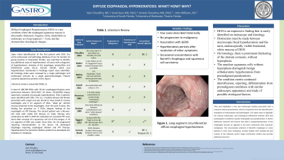
Case Description/Methods: Upon initial identification of the first patient with DEH, the entire endoscopic and pathology database of our 55 member GI group practice in Clearwater Florida, was searched to identify any additional cases of hyperkeratosis. All cases with a diagnosis of hyperkeratosis, diseases of the esophagus unspecified, and ICD9/ICD10 codes K22.9, K22.89, 530.89, 530.9 and hyperkeratosis mentioned in histology report were identified. All histology slides were reviewed by a single pathologist and endoscopic pictures by a single gastroenterologist. Patient consents obtained to present in this report. Literature review is presented (Table 1).
A total of 188,788 EGDs with 33,411 esophageal biopsies were performed between 2010-2023. Of these, 231(0.69%) biopsy specimens revealed microscopic hyperkeratosis. Only 2 patients were identified with DEH. The 1st, a healthy 60-year-old female presented with cough and was found to have Grade D erosive esophagitis and 4 cm segment of thick, “piled up” whitish mucosa proximal to the esophagitis. Over the past 9 years, this finding has persisted on 3 EGDs despite healing of the esophagitis on PPI therapy. The second patient was a 66-year-old male with stable HIV disease on triple therapy who underwent an EGD in 2019 for evaluation of a positive PET scan done after removal of a squamous cell CA of the tongue. A 10 cm segment of DEH was noted. Since then, his CA progressed requiring chemoradiation to the larynx. He developed dysphagia requiring esophageal dilation and PPI therapy. Hyperkeratosis has persisted. Neither patient has developed any dysplasia or neoplasia.
Discussion: DEH is an impressive finding that is easily identified on endoscopy and histology. Distinction must be made between microscopic/focal hyperkeratosis and the rarer, endoscopically visible thickened, white mucosa of DEH. On histology, there is prominent thickening of the stratum corneum, without hyperplasia. The anuclear squamous cells without hyperplasia distinguish benign orthokeratotic hyperkeratosis from premalignant parakeratosis. The condition merits continued identification, reporting, differentiation from premalignant conditions with similar endoscopic appearance and study of possible etiological factors.

Disclosures:
Veer Choudhry, MS1, Vivek Kaul, MD, FACG2, Umesh Choudhry, MD, FACG3, John Williams, DO3. P1899 - Diffuse Esophageal Hyperkeratosis: What? How? Why?, ACG 2023 Annual Scientific Meeting Abstracts. Vancouver, BC, Canada: American College of Gastroenterology.
Veer Choudhry, MS1, Vivek Kaul, MD, FACG2, Umesh Choudhry, MD, FACG3, John Williams, DO3
1University of South Florida, Tampa, FL; 2University of Rochester, Rochester, NY; 3Gastro Florida, Clearwater, FL
Introduction: Diffuse Esophageal Hyperkeratosis (DEH) is a rare condition where the esophageal squamous mucosa is abnormally thickened, irregular, white, identifiable on endoscopy as a long-segment ( > 3cm), and has characteristic histological changes.
Case Description/Methods: Upon initial identification of the first patient with DEH, the entire endoscopic and pathology database of our 55 member GI group practice in Clearwater Florida, was searched to identify any additional cases of hyperkeratosis. All cases with a diagnosis of hyperkeratosis, diseases of the esophagus unspecified, and ICD9/ICD10 codes K22.9, K22.89, 530.89, 530.9 and hyperkeratosis mentioned in histology report were identified. All histology slides were reviewed by a single pathologist and endoscopic pictures by a single gastroenterologist. Patient consents obtained to present in this report. Literature review is presented (Table 1).
A total of 188,788 EGDs with 33,411 esophageal biopsies were performed between 2010-2023. Of these, 231(0.69%) biopsy specimens revealed microscopic hyperkeratosis. Only 2 patients were identified with DEH. The 1st, a healthy 60-year-old female presented with cough and was found to have Grade D erosive esophagitis and 4 cm segment of thick, “piled up” whitish mucosa proximal to the esophagitis. Over the past 9 years, this finding has persisted on 3 EGDs despite healing of the esophagitis on PPI therapy. The second patient was a 66-year-old male with stable HIV disease on triple therapy who underwent an EGD in 2019 for evaluation of a positive PET scan done after removal of a squamous cell CA of the tongue. A 10 cm segment of DEH was noted. Since then, his CA progressed requiring chemoradiation to the larynx. He developed dysphagia requiring esophageal dilation and PPI therapy. Hyperkeratosis has persisted. Neither patient has developed any dysplasia or neoplasia.
Discussion: DEH is an impressive finding that is easily identified on endoscopy and histology. Distinction must be made between microscopic/focal hyperkeratosis and the rarer, endoscopically visible thickened, white mucosa of DEH. On histology, there is prominent thickening of the stratum corneum, without hyperplasia. The anuclear squamous cells without hyperplasia distinguish benign orthokeratotic hyperkeratosis from premalignant parakeratosis. The condition merits continued identification, reporting, differentiation from premalignant conditions with similar endoscopic appearance and study of possible etiological factors.

Figure: Long-segment circumferential diffuse esophageal hyperkeratosis
Disclosures:
Veer Choudhry indicated no relevant financial relationships.
Vivek Kaul: CASTLE BIOSCIENCES – Consultant. CDX DIAGNOSTICS – Consultant. COOK MEDICAL – Consultant. MOTUS GI – Consultant. STERIS – Consultant.
Umesh Choudhry indicated no relevant financial relationships.
John Williams indicated no relevant financial relationships.
Veer Choudhry, MS1, Vivek Kaul, MD, FACG2, Umesh Choudhry, MD, FACG3, John Williams, DO3. P1899 - Diffuse Esophageal Hyperkeratosis: What? How? Why?, ACG 2023 Annual Scientific Meeting Abstracts. Vancouver, BC, Canada: American College of Gastroenterology.

