Monday Poster Session
Category: Esophagus
P1903 - Esophageal Stenting in a Proximal Esophageal Perforation After Failed Surgical Repair: A Case Report
Monday, October 23, 2023
10:30 AM - 4:15 PM PT
Location: Exhibit Hall

Has Audio

Collin D. Henry, MD
University of Tennessee Health Science Center
Memphis, TN
Presenting Author(s)
Collin D.. Henry, MD1, Louis Yn, MD1, Thomas B.. Wells, DO, MBA1, William F.. Alexander, MD2
1University of Tennessee Health Science Center, Memphis, TN; 2Regional One Health, Memphis, TN
Introduction: Gunshot wounds account for 70 to 80% of traumatic esophageal perforations from penetrating trauma which results in a high morbidity and mortality. Successful management of these injuries are typically achieved by prompt surgical repair. In rare cases, esophagectomy with diversion is pursued for irreparably damaged esophagi. We present a case of a young, healthy patient with proximal esophageal perforation from a gunshot wound that was initially treated with surgical repair, but continued to have esophageal leak post-op. The esophageal leak was successfully treated by esophageal stenting negating the need for esophagostomy and likely esophagectomy.
Case Description/Methods: A 28-year-old male presented with 3 gunshot wounds. On CT, one bullet traveled through the left axilla causing injury to the esophagus. He was taken for emergent surgery. An esophagoscopy showed a large 5 cm posterior esophageal rupture 25cm from the incisors. This was repaired surgically with a right neck incision, primary surgical repair, and muscle flap. He remained in the trauma ICU for recovery. On post-op day 10, an esophagram showed active extravasation from the mid esophagus consistent with ongoing, large esophageal leak. To avoid esophagostomy followed by esophagectomy, thoracic surgery consulted the gastroenterology service for feasibility of esophageal stent. Patient underwent EGD with visualization of a large perforation only 3 cm from the upper esophageal sphincter. A Merit EndoMAXX 23mm x150mm esophageal stent was advanced and placed under fluoroscopic and endoscopic visualization with the upper end of the stent approximately 1.5 cm from the upper esophageal sphincter. Repeat esophagram showed no extravasation of contrast. Patient continued to improve and discharged home. He returned 9 weeks later for repeat EGD and stent removal. The area of prior perforation demonstrated complete healing and same day esophagram was without leak. He continues to do well and tolerate a full diet.
Discussion: The mainstay treatment for traumatic esophageal perforations remains primary surgical repair. This case demonstrates that esophageal stent is an option after failed primary surgical repair, but also for proximal perforations close to the UES. In our case, this endoscopic intervention saved a young, healthy patient from having an extensive, life-altering surgery. Our purpose is to highlight a case for the GI community that proximal esophageal stenting for traumatic perforations is an option in otherwise stable patients.

Disclosures:
Collin D.. Henry, MD1, Louis Yn, MD1, Thomas B.. Wells, DO, MBA1, William F.. Alexander, MD2. P1903 - Esophageal Stenting in a Proximal Esophageal Perforation After Failed Surgical Repair: A Case Report, ACG 2023 Annual Scientific Meeting Abstracts. Vancouver, BC, Canada: American College of Gastroenterology.
1University of Tennessee Health Science Center, Memphis, TN; 2Regional One Health, Memphis, TN
Introduction: Gunshot wounds account for 70 to 80% of traumatic esophageal perforations from penetrating trauma which results in a high morbidity and mortality. Successful management of these injuries are typically achieved by prompt surgical repair. In rare cases, esophagectomy with diversion is pursued for irreparably damaged esophagi. We present a case of a young, healthy patient with proximal esophageal perforation from a gunshot wound that was initially treated with surgical repair, but continued to have esophageal leak post-op. The esophageal leak was successfully treated by esophageal stenting negating the need for esophagostomy and likely esophagectomy.
Case Description/Methods: A 28-year-old male presented with 3 gunshot wounds. On CT, one bullet traveled through the left axilla causing injury to the esophagus. He was taken for emergent surgery. An esophagoscopy showed a large 5 cm posterior esophageal rupture 25cm from the incisors. This was repaired surgically with a right neck incision, primary surgical repair, and muscle flap. He remained in the trauma ICU for recovery. On post-op day 10, an esophagram showed active extravasation from the mid esophagus consistent with ongoing, large esophageal leak. To avoid esophagostomy followed by esophagectomy, thoracic surgery consulted the gastroenterology service for feasibility of esophageal stent. Patient underwent EGD with visualization of a large perforation only 3 cm from the upper esophageal sphincter. A Merit EndoMAXX 23mm x150mm esophageal stent was advanced and placed under fluoroscopic and endoscopic visualization with the upper end of the stent approximately 1.5 cm from the upper esophageal sphincter. Repeat esophagram showed no extravasation of contrast. Patient continued to improve and discharged home. He returned 9 weeks later for repeat EGD and stent removal. The area of prior perforation demonstrated complete healing and same day esophagram was without leak. He continues to do well and tolerate a full diet.
Discussion: The mainstay treatment for traumatic esophageal perforations remains primary surgical repair. This case demonstrates that esophageal stent is an option after failed primary surgical repair, but also for proximal perforations close to the UES. In our case, this endoscopic intervention saved a young, healthy patient from having an extensive, life-altering surgery. Our purpose is to highlight a case for the GI community that proximal esophageal stenting for traumatic perforations is an option in otherwise stable patients.

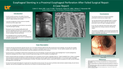
Figure: Ten days post-operative esophagram displaying continued proximal esophageal leak bilaterally prior to esophageal stenting.
Disclosures:
Collin Henry indicated no relevant financial relationships.
Louis Yn indicated no relevant financial relationships.
Thomas Wells indicated no relevant financial relationships.
William Alexander indicated no relevant financial relationships.
Collin D.. Henry, MD1, Louis Yn, MD1, Thomas B.. Wells, DO, MBA1, William F.. Alexander, MD2. P1903 - Esophageal Stenting in a Proximal Esophageal Perforation After Failed Surgical Repair: A Case Report, ACG 2023 Annual Scientific Meeting Abstracts. Vancouver, BC, Canada: American College of Gastroenterology.
