Monday Poster Session
Category: Colon
P1716 - Revealing the Unusual: Anaplastic Variant of Anorectal Kaposi Sarcoma
Monday, October 23, 2023
10:30 AM - 4:15 PM PT
Location: Exhibit Hall

Has Audio

Nikitha Chellapuram, MD
Centinela Hospital Medical Center
Los Angeles, CA
Presenting Author(s)
Nikitha Chellapuram, MD1, Rewanth Katamreddy, MD2, Saisree Reddy Adla Jala, MD3, Lincoln Luk, MD3
1Centinela Hospital Medical Center, Los Angeles, CA; 2Saint Michael’s Medical Center, Newark, NJ; 3Centinela Hospital Medical Center, Inglewood, CA
Introduction: Kaposi Sarcoma(KS) is a vascular tumor linked to Human Herpesvirus 8 (HHV-8). It has four commonly identified types associated with HHV-8. We present a case report of a patient with AIDS-related or epidemic Kaposi Sarcoma, which can vary from an incidental finding to a rapidly progressing neoplasm. While it usually manifests as a skin disease, untreated Kaposi Sarcoma can spread to other organs. In rare instances, lower gastrointestinal Kaposi Sarcoma may be found without cutaneous involvement.
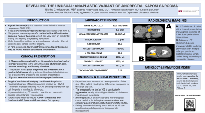
Case Description/Methods: A 29-year-old man with HIV and inconsistent antiretroviral therapy presented to the ED with severe abdominal pain, nausea, vomiting, and bloody diarrhea. The patient had a prior diagnosis and treatment for a perirectal abscess, along with multiple hospital admissions for a few months preceding the current presentation. Physical examination revealed a large perianal mass. CT scan showed suspicious neoplasm and enlarged pelvic and inguinal lymph nodes. Surgical excision with biopsy confirmed Anaplastic histological variant of Kaposi sarcoma positive for HHV-8. Immunohistochemistry results were positive for HHV-8 LNA-1 antibody, as well as the lymphatic endothelial cell markers CD34, factor VIII, and PECAM-1. Treatment included initiating HAART and outpatient follow-up, but the patient was lost to follow-up. Upon readmission, CT revealed proctitis and metastatic disease with enlarged lymph nodes. Efforts were made to ensure HAART adherence and treatment with liposomal Doxorubicin (six cycles).
Discussion: Kaposi sarcoma lesions that develop outside of the skin often exhibit histological differences compared to those on the skin. The anaplastic variant of KS is particularly aggressive locally, with a higher likelihood of deeper invasion and metastasis. Histologically, it displays variable morphological characteristics, including increased nuclear and cellular pleomorphism and a higher mitotic index. Failing to correctly identify such lesions as KS can result in delayed diagnosis or inappropriate management.

Disclosures:
Nikitha Chellapuram, MD1, Rewanth Katamreddy, MD2, Saisree Reddy Adla Jala, MD3, Lincoln Luk, MD3. P1716 - Revealing the Unusual: Anaplastic Variant of Anorectal Kaposi Sarcoma, ACG 2023 Annual Scientific Meeting Abstracts. Vancouver, BC, Canada: American College of Gastroenterology.
1Centinela Hospital Medical Center, Los Angeles, CA; 2Saint Michael’s Medical Center, Newark, NJ; 3Centinela Hospital Medical Center, Inglewood, CA
Introduction: Kaposi Sarcoma(KS) is a vascular tumor linked to Human Herpesvirus 8 (HHV-8). It has four commonly identified types associated with HHV-8. We present a case report of a patient with AIDS-related or epidemic Kaposi Sarcoma, which can vary from an incidental finding to a rapidly progressing neoplasm. While it usually manifests as a skin disease, untreated Kaposi Sarcoma can spread to other organs. In rare instances, lower gastrointestinal Kaposi Sarcoma may be found without cutaneous involvement.
Case Description/Methods: A 29-year-old man with HIV and inconsistent antiretroviral therapy presented to the ED with severe abdominal pain, nausea, vomiting, and bloody diarrhea. The patient had a prior diagnosis and treatment for a perirectal abscess, along with multiple hospital admissions for a few months preceding the current presentation. Physical examination revealed a large perianal mass. CT scan showed suspicious neoplasm and enlarged pelvic and inguinal lymph nodes. Surgical excision with biopsy confirmed Anaplastic histological variant of Kaposi sarcoma positive for HHV-8. Immunohistochemistry results were positive for HHV-8 LNA-1 antibody, as well as the lymphatic endothelial cell markers CD34, factor VIII, and PECAM-1. Treatment included initiating HAART and outpatient follow-up, but the patient was lost to follow-up. Upon readmission, CT revealed proctitis and metastatic disease with enlarged lymph nodes. Efforts were made to ensure HAART adherence and treatment with liposomal Doxorubicin (six cycles).
Discussion: Kaposi sarcoma lesions that develop outside of the skin often exhibit histological differences compared to those on the skin. The anaplastic variant of KS is particularly aggressive locally, with a higher likelihood of deeper invasion and metastasis. Histologically, it displays variable morphological characteristics, including increased nuclear and cellular pleomorphism and a higher mitotic index. Failing to correctly identify such lesions as KS can result in delayed diagnosis or inappropriate management.

Figure: A : CT abdomen&pelvis at the time of presentation showing the evidence of 6.4x4.8cm perianal soft tissue mass.
B: Follow-up CT abdomen&pelvis showing notable evidence of Proctitis with multiple enlarged pelvis and bilateral inguinal lymph nodes,likely secondary to metastatic disease.
B: Follow-up CT abdomen&pelvis showing notable evidence of Proctitis with multiple enlarged pelvis and bilateral inguinal lymph nodes,likely secondary to metastatic disease.
Disclosures:
Nikitha Chellapuram indicated no relevant financial relationships.
Rewanth Katamreddy indicated no relevant financial relationships.
Saisree Reddy Adla Jala indicated no relevant financial relationships.
Lincoln Luk indicated no relevant financial relationships.
Nikitha Chellapuram, MD1, Rewanth Katamreddy, MD2, Saisree Reddy Adla Jala, MD3, Lincoln Luk, MD3. P1716 - Revealing the Unusual: Anaplastic Variant of Anorectal Kaposi Sarcoma, ACG 2023 Annual Scientific Meeting Abstracts. Vancouver, BC, Canada: American College of Gastroenterology.
