Monday Poster Session
Category: Colon
P1723 - A Rare Case of Poorly Differentiated Medullary Carcinoma of the Colon: A Diagnostic Challenge
Monday, October 23, 2023
10:30 AM - 4:15 PM PT
Location: Exhibit Hall

Has Audio

Farhan Azad, DO
University at Buffalo
Buffalo, New York
Presenting Author(s)
Farhan Azad, DO, Clive J. Miranda, DO, MS, Alexander M. Carlson, DO, Prutha Patel, DO, Ross R. Moyer, DO, Muddasir Ayaz, MD, Bebu Ram, MD
University at Buffalo, Buffalo, NY
Introduction: The most common colon cancer is adenocarcinoma (AC). Medullary carcinoma (MC) is seen in 5 to 8 cases for every 10,000 colon cancers. Its features include female predominance, proximal location, poorly differentiated or undifferentiated morphology, microsatellite instability (MSI), and better prognosis than AC. It shares similarities to undifferentiated AC and neuroendocrine tumors (NET), making its diagnosis difficult.
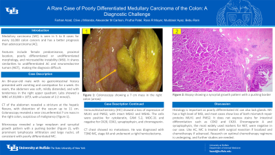
Case Description/Methods: An 84-year-old male with no prior gastrointestinal history presented with worsening constipation for a week. He noted intermittent episodes of vomiting, and his last bowel movement was 3 days prior. On exam, the abdomen was soft and mildly distended with tenderness in the right upper quadrant. Laboratory values revealed a WBC of 20,000 × 109/L with a lactate of 2.2 mmol/L. CT of the abdomen revealed a stricture at the hepatic flexure, with distention of the cecum up to 11 cm. Colonoscopy revealed a near circumferential 7 cm mass in the right colon, suspicious of malignancy (Figure 1). Microscopic examination revealed a large neoplasm and syncytial growth pattern with a pushing border (Figure 2). It also showed prominent lymphocyte infiltration (Figure 3) and large nuclei with prominent nucleoli (Figure 4), features consistent with poorly differentiated MC. The regional lymph nodes, margins, and appendix were not involved by the tumor. Immunohistochemistry (IHC) revealed loss of nuclear expression of MLH1 and PMS2, with intact MSH2 and MSH6. The tumor cells were positive for pan-cytokeratin, CAM 5.2, and MOC-31 and negative for CK20, CDX2, synaptophysin, and chromogranin. CT chest showed no evidence of metastases. He was diagnosed with T3N0 MC of the colon, stage IIA, and no adjuvant chemotherapy was given. He underwent a right hemicolectomy and was eventually discharged.
Discussion: IHC is critical in diagnosing MC as poorly differentiated AC can also lack a glandular structure. MC has a high level of MSI, and most cases show loss of both mismatch repair proteins MLH1 and PMS2, as seen here. It also typically does not express stains for intestinal differentiation such as CDX2 and CK20. Diagnosis is further complicated by its similarity to NET. Chromogranin A and synaptophysin, the most widely used markers for NET, were negative in our case. Similar to AC, MC is treated with surgical resection if localized and with chemotherapy if advanced. Research on optimal chemotherapy regimens is undergoing, and further studies are needed for this rare entity.

Disclosures:
Farhan Azad, DO, Clive J. Miranda, DO, MS, Alexander M. Carlson, DO, Prutha Patel, DO, Ross R. Moyer, DO, Muddasir Ayaz, MD, Bebu Ram, MD. P1723 - A Rare Case of Poorly Differentiated Medullary Carcinoma of the Colon: A Diagnostic Challenge, ACG 2023 Annual Scientific Meeting Abstracts. Vancouver, BC, Canada: American College of Gastroenterology.
University at Buffalo, Buffalo, NY
Introduction: The most common colon cancer is adenocarcinoma (AC). Medullary carcinoma (MC) is seen in 5 to 8 cases for every 10,000 colon cancers. Its features include female predominance, proximal location, poorly differentiated or undifferentiated morphology, microsatellite instability (MSI), and better prognosis than AC. It shares similarities to undifferentiated AC and neuroendocrine tumors (NET), making its diagnosis difficult.
Case Description/Methods: An 84-year-old male with no prior gastrointestinal history presented with worsening constipation for a week. He noted intermittent episodes of vomiting, and his last bowel movement was 3 days prior. On exam, the abdomen was soft and mildly distended with tenderness in the right upper quadrant. Laboratory values revealed a WBC of 20,000 × 109/L with a lactate of 2.2 mmol/L. CT of the abdomen revealed a stricture at the hepatic flexure, with distention of the cecum up to 11 cm. Colonoscopy revealed a near circumferential 7 cm mass in the right colon, suspicious of malignancy (Figure 1). Microscopic examination revealed a large neoplasm and syncytial growth pattern with a pushing border (Figure 2). It also showed prominent lymphocyte infiltration (Figure 3) and large nuclei with prominent nucleoli (Figure 4), features consistent with poorly differentiated MC. The regional lymph nodes, margins, and appendix were not involved by the tumor. Immunohistochemistry (IHC) revealed loss of nuclear expression of MLH1 and PMS2, with intact MSH2 and MSH6. The tumor cells were positive for pan-cytokeratin, CAM 5.2, and MOC-31 and negative for CK20, CDX2, synaptophysin, and chromogranin. CT chest showed no evidence of metastases. He was diagnosed with T3N0 MC of the colon, stage IIA, and no adjuvant chemotherapy was given. He underwent a right hemicolectomy and was eventually discharged.
Discussion: IHC is critical in diagnosing MC as poorly differentiated AC can also lack a glandular structure. MC has a high level of MSI, and most cases show loss of both mismatch repair proteins MLH1 and PMS2, as seen here. It also typically does not express stains for intestinal differentiation such as CDX2 and CK20. Diagnosis is further complicated by its similarity to NET. Chromogranin A and synaptophysin, the most widely used markers for NET, were negative in our case. Similar to AC, MC is treated with surgical resection if localized and with chemotherapy if advanced. Research on optimal chemotherapy regimens is undergoing, and further studies are needed for this rare entity.

Figure: Figure 1: Colonoscopy showing a near circumferential 7 cm mass in the right colon (arrow)
Figure 2: Microscopy showing large neoplasm with solid and syncytial growth pattern showing a pushing border
Figure 3: Microscopy showing prominent lymphocyte infiltration
Figure 4: Microscopy showing large nuclei with prominent nucleoli
Figure 2: Microscopy showing large neoplasm with solid and syncytial growth pattern showing a pushing border
Figure 3: Microscopy showing prominent lymphocyte infiltration
Figure 4: Microscopy showing large nuclei with prominent nucleoli
Disclosures:
Farhan Azad indicated no relevant financial relationships.
Clive Miranda indicated no relevant financial relationships.
Alexander Carlson indicated no relevant financial relationships.
Prutha Patel indicated no relevant financial relationships.
Ross Moyer indicated no relevant financial relationships.
Muddasir Ayaz indicated no relevant financial relationships.
Bebu Ram indicated no relevant financial relationships.
Farhan Azad, DO, Clive J. Miranda, DO, MS, Alexander M. Carlson, DO, Prutha Patel, DO, Ross R. Moyer, DO, Muddasir Ayaz, MD, Bebu Ram, MD. P1723 - A Rare Case of Poorly Differentiated Medullary Carcinoma of the Colon: A Diagnostic Challenge, ACG 2023 Annual Scientific Meeting Abstracts. Vancouver, BC, Canada: American College of Gastroenterology.
