Monday Poster Session
Category: Colon
P1729 - Metastatic Prostate Cancer Masquerading as Rectal Tumor
Monday, October 23, 2023
10:30 AM - 4:15 PM PT
Location: Exhibit Hall

Has Audio

Justice Arhinful, MD
Howard University Hospital
Washington, DC
Presenting Author(s)
Justice Arhinful, MD, Suryanarayana Reddy Challa, MD, Philip Oppong-Twene, MD, MPH, Getachew Mekasha, MD, PhD, Laiyemo Adeyinka, MD, MPH, FACG
Howard University Hospital, Washington, DC
Introduction: Prostate cancer is the second most common cancer among men worldwide. At the time of diagnosis, more than 78 percent of patients have localized disease while 12 and 6 percent of patients have regional lymph node involvement and distant metastases respectively. Common sites of metastasis include bone, lung and liver. However, in spite of its proximity to the prostate, rectal involvement of metastatic prostate cancer is rare. We present a case of an abnormal extension of a prostate cancer into the rectum.
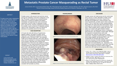
Case Description/Methods: A 74-year-old male with history of hypertension, diabetes mellitus, and stage 4 prostate cancer diagnosed in 2014 following complaints of urinary symptoms with a PSA level of 66.9. He underwent TURP but he declined treatment until 5 years later at which time he underwent an ultrasound-guided biopsy of the prostate. Pathology was consistent with adenocarcinoma. Bone scan was negative for distant metastasis. CT scan abdomen/pelvis showed multiple retroperitoneal lymph nodes and a right pericolonic pelvic lymph node abutting the sigmoid colon. Colonoscopy in 2019 showed rectal hyperplastic polyp (image A). He completed radiation therapy after which he was lost to follow-up. Three years later, CT scan showed extensive peritoneal and omental masses, L3 & L5 vertebral bodies blastic lesions and multiple left pulmonary nodules concerning for ongoing metastatic prostate cancer. Diagnostic colonoscopy done in 2023 due hematochezia and changes in bowel habit showed interval development of a partially obstructing large frond-like mass in the rectum (image B). Biopsy confirmed poorly differentiated adenocarcinoma consistent with prostate as primary.
Discussion: Metastatic prostate cancer usually metastasizes to the bone, lung and liver. In spite of the prostate's close proximity to the rectum, rectal involvement is rare, in part due to the Denonvilliers fascia acting as critical barrier to local extension of the tumor. Although the diagnosis of prostate cancer with rectal involvement can easily be made based on rectal examination, radiologic characteristics and histology of rectal biopsy together with elevated PSA, it can occasionally be confused with primary rectal carcinoma, because of similar radiologic appearances and clinical presentations such as large-bowel obstruction and rectal bleeding, etc., especially in the setting of a normal colonoscopy 3 years prior. Accurate and prompt diagnosis of these 2 distinct entities is crucial since the treatment and prognosis differ widely.

Disclosures:
Justice Arhinful, MD, Suryanarayana Reddy Challa, MD, Philip Oppong-Twene, MD, MPH, Getachew Mekasha, MD, PhD, Laiyemo Adeyinka, MD, MPH, FACG. P1729 - Metastatic Prostate Cancer Masquerading as Rectal Tumor, ACG 2023 Annual Scientific Meeting Abstracts. Vancouver, BC, Canada: American College of Gastroenterology.
Howard University Hospital, Washington, DC
Introduction: Prostate cancer is the second most common cancer among men worldwide. At the time of diagnosis, more than 78 percent of patients have localized disease while 12 and 6 percent of patients have regional lymph node involvement and distant metastases respectively. Common sites of metastasis include bone, lung and liver. However, in spite of its proximity to the prostate, rectal involvement of metastatic prostate cancer is rare. We present a case of an abnormal extension of a prostate cancer into the rectum.
Case Description/Methods: A 74-year-old male with history of hypertension, diabetes mellitus, and stage 4 prostate cancer diagnosed in 2014 following complaints of urinary symptoms with a PSA level of 66.9. He underwent TURP but he declined treatment until 5 years later at which time he underwent an ultrasound-guided biopsy of the prostate. Pathology was consistent with adenocarcinoma. Bone scan was negative for distant metastasis. CT scan abdomen/pelvis showed multiple retroperitoneal lymph nodes and a right pericolonic pelvic lymph node abutting the sigmoid colon. Colonoscopy in 2019 showed rectal hyperplastic polyp (image A). He completed radiation therapy after which he was lost to follow-up. Three years later, CT scan showed extensive peritoneal and omental masses, L3 & L5 vertebral bodies blastic lesions and multiple left pulmonary nodules concerning for ongoing metastatic prostate cancer. Diagnostic colonoscopy done in 2023 due hematochezia and changes in bowel habit showed interval development of a partially obstructing large frond-like mass in the rectum (image B). Biopsy confirmed poorly differentiated adenocarcinoma consistent with prostate as primary.
Discussion: Metastatic prostate cancer usually metastasizes to the bone, lung and liver. In spite of the prostate's close proximity to the rectum, rectal involvement is rare, in part due to the Denonvilliers fascia acting as critical barrier to local extension of the tumor. Although the diagnosis of prostate cancer with rectal involvement can easily be made based on rectal examination, radiologic characteristics and histology of rectal biopsy together with elevated PSA, it can occasionally be confused with primary rectal carcinoma, because of similar radiologic appearances and clinical presentations such as large-bowel obstruction and rectal bleeding, etc., especially in the setting of a normal colonoscopy 3 years prior. Accurate and prompt diagnosis of these 2 distinct entities is crucial since the treatment and prognosis differ widely.

Figure: Endoscopic photographs of the rectum showing hyperplastic polyp (Image A) and interval development of rectal mass (Image B)
Disclosures:
Justice Arhinful indicated no relevant financial relationships.
Suryanarayana Reddy Challa indicated no relevant financial relationships.
Philip Oppong-Twene indicated no relevant financial relationships.
Getachew Mekasha indicated no relevant financial relationships.
Laiyemo Adeyinka indicated no relevant financial relationships.
Justice Arhinful, MD, Suryanarayana Reddy Challa, MD, Philip Oppong-Twene, MD, MPH, Getachew Mekasha, MD, PhD, Laiyemo Adeyinka, MD, MPH, FACG. P1729 - Metastatic Prostate Cancer Masquerading as Rectal Tumor, ACG 2023 Annual Scientific Meeting Abstracts. Vancouver, BC, Canada: American College of Gastroenterology.
