Monday Poster Session
Category: Biliary/Pancreas
P1497 - A Diagnostically Difficult Case of Type III Choledochocele Masquerading as a Pancreatic Cystic Mass
Monday, October 23, 2023
10:30 AM - 4:15 PM PT
Location: Exhibit Hall

Has Audio

Alexander Garcia, DO
Cooper University Hospital
Camden, NJ
Presenting Author(s)
Alexander Garcia, DO1, Rita Auro, MD1, Gina Wodarczyk, BS2, Jenny Melli, MD1
1Cooper University Hospital, Camden, NJ; 2Cooper Medical School of Rowan University, Camden, NJ
Introduction: Choledochal cysts (CCs) are rare congenital cystic dilatations of the biliary tree. Type III CCs, also referred to as choledochoceles, are the rarest subtypes representing 1-4.5% of all CCs and are defined by their involvement of the distal common bile duct and duodenum. Here we present the case of a patient with a pancreatic mass that was ultimately found to be a choledochocele.
Case Description/Methods: A 56-year-old male with a history of cholecystectomy presented for evaluation of painless obstructive jaundice. For several months he had been bothered by fatigue, pruritis, and dyspnea on exertion. On exam he was noted to be prominently jaundiced. LFTs at presentation yielded the following: ALT of 140 U/L, AST of 191 U/L, ALP of 1,788 (U/L), and direct bilirubin of 9.3 mg/dL. Right upper quadrant ultrasound revealed mild dilation of the pancreatic duct at the pancreatic head and a CBD observed to be 9.6 mm. Follow-up CT showed a heterogenous cystic mass involving the pancreatic head that measured 4.0 x 3.3 cm in size. CA 19-9 was elevated at 62. MRCP was attempted, but unsuccessful due to claustrophobia. ERCP was aborted early on during the procedure as the major papilla was obscured within a food residue laden diverticulum. Following placement of a transhepatic biliary drain the patient noted significant improvement in symptoms and his LFTs began down trending. On repeat CT there was notable absence of the previously seen cystic structure at the pancreatic head raising the possibility of a choledochocele. Several months later the patient went on to have an elective Whipple procedure with biopsy confirming the presence of Type III CC.
Discussion: Choledochal cysts (CCs) are quite rare and occur in approximately 0.007% of live births in the United States. Type III CCs arise from dilation of the most distal portion of the CBD at the junction with the duodenum. Radiographically, Type III CCs appear as cystic structures abutting the medial wall of the duodenum. In our case imaging was most consistent with a discrete pancreatic lesion with no involvement of the bowel. Moreover, with an elevated CA 19-9 and painless obstructive jaundice a pancreatic neoplastic process was of concern. Our case shows the need to keep Type III CCs in the differential for patients who present with discrete pancreatic cysts/masses.

Disclosures:
Alexander Garcia, DO1, Rita Auro, MD1, Gina Wodarczyk, BS2, Jenny Melli, MD1. P1497 - A Diagnostically Difficult Case of Type III Choledochocele Masquerading as a Pancreatic Cystic Mass, ACG 2023 Annual Scientific Meeting Abstracts. Vancouver, BC, Canada: American College of Gastroenterology.
1Cooper University Hospital, Camden, NJ; 2Cooper Medical School of Rowan University, Camden, NJ
Introduction: Choledochal cysts (CCs) are rare congenital cystic dilatations of the biliary tree. Type III CCs, also referred to as choledochoceles, are the rarest subtypes representing 1-4.5% of all CCs and are defined by their involvement of the distal common bile duct and duodenum. Here we present the case of a patient with a pancreatic mass that was ultimately found to be a choledochocele.
Case Description/Methods: A 56-year-old male with a history of cholecystectomy presented for evaluation of painless obstructive jaundice. For several months he had been bothered by fatigue, pruritis, and dyspnea on exertion. On exam he was noted to be prominently jaundiced. LFTs at presentation yielded the following: ALT of 140 U/L, AST of 191 U/L, ALP of 1,788 (U/L), and direct bilirubin of 9.3 mg/dL. Right upper quadrant ultrasound revealed mild dilation of the pancreatic duct at the pancreatic head and a CBD observed to be 9.6 mm. Follow-up CT showed a heterogenous cystic mass involving the pancreatic head that measured 4.0 x 3.3 cm in size. CA 19-9 was elevated at 62. MRCP was attempted, but unsuccessful due to claustrophobia. ERCP was aborted early on during the procedure as the major papilla was obscured within a food residue laden diverticulum. Following placement of a transhepatic biliary drain the patient noted significant improvement in symptoms and his LFTs began down trending. On repeat CT there was notable absence of the previously seen cystic structure at the pancreatic head raising the possibility of a choledochocele. Several months later the patient went on to have an elective Whipple procedure with biopsy confirming the presence of Type III CC.
Discussion: Choledochal cysts (CCs) are quite rare and occur in approximately 0.007% of live births in the United States. Type III CCs arise from dilation of the most distal portion of the CBD at the junction with the duodenum. Radiographically, Type III CCs appear as cystic structures abutting the medial wall of the duodenum. In our case imaging was most consistent with a discrete pancreatic lesion with no involvement of the bowel. Moreover, with an elevated CA 19-9 and painless obstructive jaundice a pancreatic neoplastic process was of concern. Our case shows the need to keep Type III CCs in the differential for patients who present with discrete pancreatic cysts/masses.

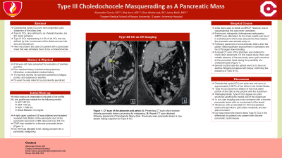
Figure: CT scan revealing a heterogeneous low-attenuation mass involving the pancreatic head (A). Following biliary drain placement, the complicated cystic structure in the head/uncinate process of the pancreas is no longer visualized (B) raising suspicion for a choledochal cyst.
Disclosures:
Alexander Garcia indicated no relevant financial relationships.
Rita Auro indicated no relevant financial relationships.
Gina Wodarczyk indicated no relevant financial relationships.
Jenny Melli indicated no relevant financial relationships.
Alexander Garcia, DO1, Rita Auro, MD1, Gina Wodarczyk, BS2, Jenny Melli, MD1. P1497 - A Diagnostically Difficult Case of Type III Choledochocele Masquerading as a Pancreatic Cystic Mass, ACG 2023 Annual Scientific Meeting Abstracts. Vancouver, BC, Canada: American College of Gastroenterology.
