Monday Poster Session
Category: Biliary/Pancreas
P1503 - Hemosuccus Pancreaticus: A Challenging Diagnosis Beyond Endoscopy
Monday, October 23, 2023
10:30 AM - 4:15 PM PT
Location: Exhibit Hall

Has Audio
- FC
Fernando Contreras, MD
Centro de Gastroenterologia Avanzada
Santo Domingo, Distrito Nacional, Dominican Republic
Presenting Author(s)
Jorge Solivey, MD1, Carlota Cabrer, MD2, Patricia Yunen, MD1, Norberto Rojas, MD1, Louis M. Morel-Ovalle, MD1, Fernando Contreras, MD3
1Centro de Diagnóstico Medicina Avanzada y Telemedicina, Santo Domingo, Distrito Nacional, Dominican Republic; 2Centro de Diagnostico Medicina Avanzada y Telemedicina, Santo Domingo, Distrito Nacional, Dominican Republic; 3Centro de Gastroenterologia Avanzada, Santo Domingo, Distrito Nacional, Dominican Republic
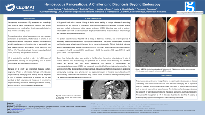
Introduction: Hemosuccus pancreaticus (HP) is a highly unusual cause of bleeding in the upper gastrointestinal tract, with arterial aneurysms being the primary cause. HP can be life-threatening due to the severity of the bleeding it causes. However, diagnosing HP is difficult due to its rarity, and only 30% of patients with endoscopy can detect active bleeding through the papilla. To identify the causative artery and guide therapeutic interventions, angiography is considered the gold standard for both diagnosis and treatment, as it helps to outline the arterial anatomy.
Case Description/Methods: A 76-year-old male with a past medical history of alcoholic pancreatitis and two episodes of gastrointestinal bleeding associated with severe anemia that were investigated with several endoscopies, VCE, and laboratories in the past without an apparent cause of the anemia presented with a history of dizziness, weakness, and several episodes of alternating melena and hematochezia. On physical examination, his skin was pale, his blood pressure levels were borderline low, his heart rate was at the upper limit of normal, and he had a positive Tilt test. Rectal examination revealed glistening red blood. Laboratories values showed: Hb 6.2 mg/dl, Hct 20%, platelets 145,000 UL, creatinine 1.30 mg/dl, BUN 35 mg/dl.
Given these findings, the patient was admitted, and 2 U of packed red blood cells were transfused. A colonoscopy was performed with no evident cause of bleeding. During the EGD, active bleeding in the second portion of the duodenum through Vater´s ampulla was seen. Interventional Radiology performed an abdominal arteriography, showing a large splenic artery and a pseudoaneurysm of the dorsal pancreatic artery with active bleeding, which was embolized with six coils. Bleeding control was achieved, and the patient recovered well.
Discussion: It is important to consider HP as a possible cause of bleeding, particularly in patients with risk factors such as chronic pancreatitis or alcohol abuse. If internal GI causes have been ruled out, it is crucial to explore other possible causes of obscure GI bleeding. When endoscopic methods are limited, alternative diagnostic methods like angiography may be necessary. The successful management of HP, in this case, shows the importance of a collaborative approach involving GI and Radiology specialists.

Disclosures:
Jorge Solivey, MD1, Carlota Cabrer, MD2, Patricia Yunen, MD1, Norberto Rojas, MD1, Louis M. Morel-Ovalle, MD1, Fernando Contreras, MD3. P1503 - Hemosuccus Pancreaticus: A Challenging Diagnosis Beyond Endoscopy, ACG 2023 Annual Scientific Meeting Abstracts. Vancouver, BC, Canada: American College of Gastroenterology.
1Centro de Diagnóstico Medicina Avanzada y Telemedicina, Santo Domingo, Distrito Nacional, Dominican Republic; 2Centro de Diagnostico Medicina Avanzada y Telemedicina, Santo Domingo, Distrito Nacional, Dominican Republic; 3Centro de Gastroenterologia Avanzada, Santo Domingo, Distrito Nacional, Dominican Republic
Introduction: Hemosuccus pancreaticus (HP) is a highly unusual cause of bleeding in the upper gastrointestinal tract, with arterial aneurysms being the primary cause. HP can be life-threatening due to the severity of the bleeding it causes. However, diagnosing HP is difficult due to its rarity, and only 30% of patients with endoscopy can detect active bleeding through the papilla. To identify the causative artery and guide therapeutic interventions, angiography is considered the gold standard for both diagnosis and treatment, as it helps to outline the arterial anatomy.
Case Description/Methods: A 76-year-old male with a past medical history of alcoholic pancreatitis and two episodes of gastrointestinal bleeding associated with severe anemia that were investigated with several endoscopies, VCE, and laboratories in the past without an apparent cause of the anemia presented with a history of dizziness, weakness, and several episodes of alternating melena and hematochezia. On physical examination, his skin was pale, his blood pressure levels were borderline low, his heart rate was at the upper limit of normal, and he had a positive Tilt test. Rectal examination revealed glistening red blood. Laboratories values showed: Hb 6.2 mg/dl, Hct 20%, platelets 145,000 UL, creatinine 1.30 mg/dl, BUN 35 mg/dl.
Given these findings, the patient was admitted, and 2 U of packed red blood cells were transfused. A colonoscopy was performed with no evident cause of bleeding. During the EGD, active bleeding in the second portion of the duodenum through Vater´s ampulla was seen. Interventional Radiology performed an abdominal arteriography, showing a large splenic artery and a pseudoaneurysm of the dorsal pancreatic artery with active bleeding, which was embolized with six coils. Bleeding control was achieved, and the patient recovered well.
Discussion: It is important to consider HP as a possible cause of bleeding, particularly in patients with risk factors such as chronic pancreatitis or alcohol abuse. If internal GI causes have been ruled out, it is crucial to explore other possible causes of obscure GI bleeding. When endoscopic methods are limited, alternative diagnostic methods like angiography may be necessary. The successful management of HP, in this case, shows the importance of a collaborative approach involving GI and Radiology specialists.

Figure: A) EGD showing blood in the second portion of the duodenum near the Vater´s ampulla
B) EGD showing active bleeding in the second portion of the duodenum through the Vater´s ampulla
C) Arteriography showing a pseudoaneurysm of the dorsal pancreatic artery with active bleeding
D) Pseudoaneurysm of the dorsal pancreatic artery embolized with a total of 6 coils.
B) EGD showing active bleeding in the second portion of the duodenum through the Vater´s ampulla
C) Arteriography showing a pseudoaneurysm of the dorsal pancreatic artery with active bleeding
D) Pseudoaneurysm of the dorsal pancreatic artery embolized with a total of 6 coils.
Disclosures:
Jorge Solivey indicated no relevant financial relationships.
Carlota Cabrer indicated no relevant financial relationships.
Patricia Yunen indicated no relevant financial relationships.
Norberto Rojas indicated no relevant financial relationships.
Louis Morel-Ovalle indicated no relevant financial relationships.
Fernando Contreras indicated no relevant financial relationships.
Jorge Solivey, MD1, Carlota Cabrer, MD2, Patricia Yunen, MD1, Norberto Rojas, MD1, Louis M. Morel-Ovalle, MD1, Fernando Contreras, MD3. P1503 - Hemosuccus Pancreaticus: A Challenging Diagnosis Beyond Endoscopy, ACG 2023 Annual Scientific Meeting Abstracts. Vancouver, BC, Canada: American College of Gastroenterology.

