Monday Poster Session
Category: Biliary/Pancreas
P1504 - Endoscopic Management of a Bronchobiliary Fistula With EUS-Guided Hepaticogastrostomy in the Setting of a Complex Postsurgical Anatomy
Monday, October 23, 2023
10:30 AM - 4:15 PM PT
Location: Exhibit Hall

Has Audio

Donna Maria Abboud, MD
Mayo Clinic
Rochester, MN
Presenting Author(s)
Award: Presidential Poster Award
Donna Maria Abboud, MD, Aliana Bofill-Garcia, MD
Mayo Clinic, Rochester, MN
Introduction: Bronchobiliary fistula (BBF) is a rare and challenging complication resulting in an abnormal connection between the bronchial system and biliary tree. Few reports have described BBF, but its management is still not clearly defined. Here, we report the successful endoscopic management of a BBF in the setting of complex postsurgical anatomy.
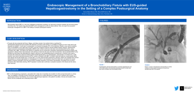
Case Description/Methods: A 75-year-old man presented with fever, fatigue, and bilious emesis. His medical history is pertinent for extrahepatic cholangiocarcinoma treated with Whipple’s procedure with findings on surveillance imaging of a liver mass which was biopsied and ablated 1 month prior to presentation. A computer tomography (CT) of the abdomen showed a new central cavitating area of consolidation in the right lower lung (RLL) that was contiguous with the recently ablated hepatic segment. The patient was started on antibiotics and an endoscopic retrograde cholangiopancreatography (ERCP) was requested to attempt biliary decompression with stenting to divert the bile away from the bronchial tree. Device-assisted ERCP using Rendezvous technique was attempted but unable to advance to the hepaticojejunostomy anastomosis. A decision was made to proceed with endoscopic ultrasound (EUS)-guided hepaticogastrostomy. On cholangiogram, extravasation of contrast from a right posterior duct branch extending towards the RLL cavity is consistent with BBF (figure 1a). A covered metal stent with two coaxial plastic stents were placed into the left hepatic duct. A month later, CT of the chest showed resolved RLL abscess. ERCP was repeated with minimal extravasation of contrast appreciated on cholangiogram for which it was decided to leave the metal stent and upsize the coaxial pigtails to enhance drainage of the liver and achieve complete resolution of the fistula. Nevertheless, the patient had no further symptoms after initial biliary stenting. ERCP repeated 3 months later confirmed a complete resolution of the fistula (figure 1b) and the stents were removed.
Discussion: BBF is a life‐threatening complication, associated with a high rate of morbidity and mortality for which prompt intervention is critical. Here, we highlight a minimally invasive endoscopic approach as a viable alternative to surgery even in patients with complex postsurgical anatomy. Nonetheless, the success of the technique depends on the patient’s condition, the size and location of the fistula, and the complexity and underlying cause of BBF.

Disclosures:
Donna Maria Abboud, MD, Aliana Bofill-Garcia, MD. P1504 - Endoscopic Management of a Bronchobiliary Fistula With EUS-Guided Hepaticogastrostomy in the Setting of a Complex Postsurgical Anatomy, ACG 2023 Annual Scientific Meeting Abstracts. Vancouver, BC, Canada: American College of Gastroenterology.
Donna Maria Abboud, MD, Aliana Bofill-Garcia, MD
Mayo Clinic, Rochester, MN
Introduction: Bronchobiliary fistula (BBF) is a rare and challenging complication resulting in an abnormal connection between the bronchial system and biliary tree. Few reports have described BBF, but its management is still not clearly defined. Here, we report the successful endoscopic management of a BBF in the setting of complex postsurgical anatomy.
Case Description/Methods: A 75-year-old man presented with fever, fatigue, and bilious emesis. His medical history is pertinent for extrahepatic cholangiocarcinoma treated with Whipple’s procedure with findings on surveillance imaging of a liver mass which was biopsied and ablated 1 month prior to presentation. A computer tomography (CT) of the abdomen showed a new central cavitating area of consolidation in the right lower lung (RLL) that was contiguous with the recently ablated hepatic segment. The patient was started on antibiotics and an endoscopic retrograde cholangiopancreatography (ERCP) was requested to attempt biliary decompression with stenting to divert the bile away from the bronchial tree. Device-assisted ERCP using Rendezvous technique was attempted but unable to advance to the hepaticojejunostomy anastomosis. A decision was made to proceed with endoscopic ultrasound (EUS)-guided hepaticogastrostomy. On cholangiogram, extravasation of contrast from a right posterior duct branch extending towards the RLL cavity is consistent with BBF (figure 1a). A covered metal stent with two coaxial plastic stents were placed into the left hepatic duct. A month later, CT of the chest showed resolved RLL abscess. ERCP was repeated with minimal extravasation of contrast appreciated on cholangiogram for which it was decided to leave the metal stent and upsize the coaxial pigtails to enhance drainage of the liver and achieve complete resolution of the fistula. Nevertheless, the patient had no further symptoms after initial biliary stenting. ERCP repeated 3 months later confirmed a complete resolution of the fistula (figure 1b) and the stents were removed.
Discussion: BBF is a life‐threatening complication, associated with a high rate of morbidity and mortality for which prompt intervention is critical. Here, we highlight a minimally invasive endoscopic approach as a viable alternative to surgery even in patients with complex postsurgical anatomy. Nonetheless, the success of the technique depends on the patient’s condition, the size and location of the fistula, and the complexity and underlying cause of BBF.

Figure: Figure 1a: Cholangiogram showing extravasation of contrast originating from one of the right posterior duct branches extending towards the right lower lobe cavity consistent with bronchobiliary fistula.
Figure 1b: Balloon occlusion cholangiogram demonstrating no further evidence of extravasation of contrast at the previous identified leak site on final ERCP.
Figure 1b: Balloon occlusion cholangiogram demonstrating no further evidence of extravasation of contrast at the previous identified leak site on final ERCP.
Disclosures:
Donna Maria Abboud indicated no relevant financial relationships.
Aliana Bofill-Garcia indicated no relevant financial relationships.
Donna Maria Abboud, MD, Aliana Bofill-Garcia, MD. P1504 - Endoscopic Management of a Bronchobiliary Fistula With EUS-Guided Hepaticogastrostomy in the Setting of a Complex Postsurgical Anatomy, ACG 2023 Annual Scientific Meeting Abstracts. Vancouver, BC, Canada: American College of Gastroenterology.

