Tuesday Poster Session
Category: IBD
P3667 - Inflammatory Pseudotumor of the Liver: A Diagnostic Challenge and Uncommon Association With Crohn's Disease
Tuesday, October 24, 2023
10:30 AM - 4:00 PM PT
Location: Exhibit Hall

- SS
Saher Sheikh, MD
Nassau University Medical Center
East Meadow, NY
Presenting Author(s)
Saher Sheikh, MD1, Ana P. Rivera Arauz, MD1, Dhir Gala, BS2, Jasmine Tsai, BS3, Sandra Gomez, MD1, Sunny Qi-Huang, MD1, Paul Mustacchia, MD, MBA1
1Nassau University Medical Center, East Meadow, NY; 2American University of the Caribbean School of Medicine, Plainview, NY; 3American University of the Caribbean School of Medicine, East Meadow, NY
Introduction: Liver pseudotumors, which resemble malignant tumors in clinical and radiological aspects, include a rare subtype called inflammatory pseudotumor (IPT) which is characterized by chronic inflammation. Although IPTs can develop in different organs, their connection to Crohn's disease is exceptionally uncommon.
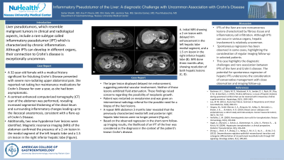
Case Description/Methods: A 32-year-old female with a medical history significant for fistulizing Crohn's Disease presented with severe non-radiating upper abdominal pain. She reported not taking her maintenance medications for Crohn's Disease for over a year, as she had been asymptomatic. A contrast-enhanced computerized tomography (CT) scan of the abdomen was performed, revealing increased segmental thickening of the distal ileum and surrounding inflammatory changes leading up to the ileocecal anastomosis, consistent with a flare-up of Crohn's Disease. Additionally, two new hypodense liver lesions were identified. Magnetic resonance imaging (MRI) of the abdomen confirmed the presence of a 2 cm lesion in the medial segment of the left hepatic lobe and a 1.5 cm lesion in the right inferior hepatic lobe (Figure). The larger lesion displayed delayed rim enhancement, suggesting potential vascular involvement. Neither of these lesions exhibited fluid attenuation. These findings raised concerns regarding the possibility of neoplastic growth. She was restarted on mesalamine and was given an interventional radiology referral for the possible need for a biopsy of the liver lesions. A repeat MRI abdomen 3 months later revealed that the previously characterized medial left and posterior right hepatic lobe lesions were no longer present (Figure). Based on the observed regression in the short-term follow-up imaging results, the likelihood of an IPT of the liver was considered as the diagnosis in the context of the patient's known Crohn's disease.
Discussion: IPTs of the liver are rare noncancerous lesions characterized by fibrous tissue and inflammatory cell infiltration. Although IPTs can occur in various organs, hepatic involvement is relatively uncommon. Spontaneous regression has been observed in some cases, highlighting the consideration of regular imaging follow-up in selected patients. This case highlights the diagnostic challenges and rare association between IPTs of the liver and Crohn's disease. The potential for spontaneous regression of hepatic IPTs underscores the consideration of conservative management with close observation and imaging follow-up.

Disclosures:
Saher Sheikh, MD1, Ana P. Rivera Arauz, MD1, Dhir Gala, BS2, Jasmine Tsai, BS3, Sandra Gomez, MD1, Sunny Qi-Huang, MD1, Paul Mustacchia, MD, MBA1. P3667 - Inflammatory Pseudotumor of the Liver: A Diagnostic Challenge and Uncommon Association With Crohn's Disease, ACG 2023 Annual Scientific Meeting Abstracts. Vancouver, BC, Canada: American College of Gastroenterology.
1Nassau University Medical Center, East Meadow, NY; 2American University of the Caribbean School of Medicine, Plainview, NY; 3American University of the Caribbean School of Medicine, East Meadow, NY
Introduction: Liver pseudotumors, which resemble malignant tumors in clinical and radiological aspects, include a rare subtype called inflammatory pseudotumor (IPT) which is characterized by chronic inflammation. Although IPTs can develop in different organs, their connection to Crohn's disease is exceptionally uncommon.
Case Description/Methods: A 32-year-old female with a medical history significant for fistulizing Crohn's Disease presented with severe non-radiating upper abdominal pain. She reported not taking her maintenance medications for Crohn's Disease for over a year, as she had been asymptomatic. A contrast-enhanced computerized tomography (CT) scan of the abdomen was performed, revealing increased segmental thickening of the distal ileum and surrounding inflammatory changes leading up to the ileocecal anastomosis, consistent with a flare-up of Crohn's Disease. Additionally, two new hypodense liver lesions were identified. Magnetic resonance imaging (MRI) of the abdomen confirmed the presence of a 2 cm lesion in the medial segment of the left hepatic lobe and a 1.5 cm lesion in the right inferior hepatic lobe (Figure). The larger lesion displayed delayed rim enhancement, suggesting potential vascular involvement. Neither of these lesions exhibited fluid attenuation. These findings raised concerns regarding the possibility of neoplastic growth. She was restarted on mesalamine and was given an interventional radiology referral for the possible need for a biopsy of the liver lesions. A repeat MRI abdomen 3 months later revealed that the previously characterized medial left and posterior right hepatic lobe lesions were no longer present (Figure). Based on the observed regression in the short-term follow-up imaging results, the likelihood of an IPT of the liver was considered as the diagnosis in the context of the patient's known Crohn's disease.
Discussion: IPTs of the liver are rare noncancerous lesions characterized by fibrous tissue and inflammatory cell infiltration. Although IPTs can occur in various organs, hepatic involvement is relatively uncommon. Spontaneous regression has been observed in some cases, highlighting the consideration of regular imaging follow-up in selected patients. This case highlights the diagnostic challenges and rare association between IPTs of the liver and Crohn's disease. The potential for spontaneous regression of hepatic IPTs underscores the consideration of conservative management with close observation and imaging follow-up.

Figure: A. Initial MRI showing a 2 cm lesion with delayed rim enhancement in the left hepatic lobe medial segment, and a 1.5 cm lesion in the right inferior hepatic lobe (B). MRI done three months after, showing resolution of both hepatic lesions (C, D).
Disclosures:
Saher Sheikh indicated no relevant financial relationships.
Ana Rivera Arauz indicated no relevant financial relationships.
Dhir Gala indicated no relevant financial relationships.
Jasmine Tsai indicated no relevant financial relationships.
Sandra Gomez indicated no relevant financial relationships.
Sunny Qi-Huang indicated no relevant financial relationships.
Paul Mustacchia indicated no relevant financial relationships.
Saher Sheikh, MD1, Ana P. Rivera Arauz, MD1, Dhir Gala, BS2, Jasmine Tsai, BS3, Sandra Gomez, MD1, Sunny Qi-Huang, MD1, Paul Mustacchia, MD, MBA1. P3667 - Inflammatory Pseudotumor of the Liver: A Diagnostic Challenge and Uncommon Association With Crohn's Disease, ACG 2023 Annual Scientific Meeting Abstracts. Vancouver, BC, Canada: American College of Gastroenterology.
