Tuesday Poster Session
Category: IBD
P3668 - Unveiling the Overlap: Takayasu Arteritis Manifestation in an Adult Patient With Crohn's Disease
Tuesday, October 24, 2023
10:30 AM - 4:00 PM PT
Location: Exhibit Hall

Has Audio

Abu Abbasi, MD
Loyola University Medical Center
Maywood, IL
Presenting Author(s)
Abu Fahad Abbasi, MD1, Filippa Trikantzopoulou, MD2, Joshua Amalraj, BS3, Krishna Patel, DO2, Amar Naik, MD2, Ayokunle T.. Abegunde, MD1
1Loyola University Medical Center, Maywood, IL; 2Loyola University Medical Center, Chicago, IL; 3Ross University School of Medicine, Chicago, IL
Introduction: Inflammatory bowel disease (IBD) is a chronic inflammatory condition of the gastrointestinal (GI) tract subclassified into ulcerative colitis (UC) and Crohn’s disease (CD). Takayasu’s arteritis (TAK) is a large-vessel form of vasculitis which commonly affects the aorta and main branches. Though rare, concurrence of IBD and TAK has been shown to exist in 9% of IBD cases. We report a case of newly diagnosed TAK in an established CD patient.
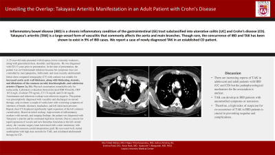
Case Description/Methods: A 25-year-old male presented with dyspnea, lower extremity weakness, along with generalized chest, shoulder, and hip pains. He was diagnosed with CD 13 years prior to presentation. At the time of presentation, the patient was on Ustekinumab infusions because his symptoms were not controlled by mercaptopurine, Infliximab, and most recently adalimumab. Initial chest computed tomography (CT) with contrast was notable for increased aortic arch wall thickness, along with thickening, stenosis, and dilatations of the common carotid, brachiocephalic, and subclavian arteries (Figures 1a, 1b). Physical examination remarkable only for tachycardia. Laboratory evaluation showed elevated ESR 95mm/hr, CRP 163.6 mg/L, D-dimer 579 ng/mL, C3 174 mg/dL and C4
40 mg/dL. Autoimmune and infectious workup were otherwise negative. The patient was presumptively diagnosed with vasculitis and discharged on steroid therapy, only to return a couple of weeks later with worsening symptoms of shortness of breath, dizziness, headaches, and left sided neck pressure. Repeat chest CT displayed significantly rapid expansion of the left common carotid artery. Based on initial workup, improvement of inflammatory markers with steroids, and imaging findings, the patient was
diagnosed with Takayasu’s arteritis and he continued high dose steroids. Due to concern for rapid expansion of vessels and new thrombus formation in the left carotid artery, the vascular surgery team intervened with a mini sternotomy with aorta-to-left common carotid interposition graft. He recovered well, started azathioprine with high dose steroids for TAK, and reinitiated adalimumab therapy for CD.
Discussion: There are increasing reports of TAK in adolescent and adult patients with IBD (UC and CD) but the pathophysiological mechanism for the association is unclear. TAK can develop in IBD patients with uncontrolled symptoms or remission. Therefore, a high index of suspicion for co-occurrence of TAK in IBD patients is crucial to preventing sequelae and complications.

Disclosures:
Abu Fahad Abbasi, MD1, Filippa Trikantzopoulou, MD2, Joshua Amalraj, BS3, Krishna Patel, DO2, Amar Naik, MD2, Ayokunle T.. Abegunde, MD1. P3668 - Unveiling the Overlap: Takayasu Arteritis Manifestation in an Adult Patient With Crohn's Disease, ACG 2023 Annual Scientific Meeting Abstracts. Vancouver, BC, Canada: American College of Gastroenterology.
1Loyola University Medical Center, Maywood, IL; 2Loyola University Medical Center, Chicago, IL; 3Ross University School of Medicine, Chicago, IL
Introduction: Inflammatory bowel disease (IBD) is a chronic inflammatory condition of the gastrointestinal (GI) tract subclassified into ulcerative colitis (UC) and Crohn’s disease (CD). Takayasu’s arteritis (TAK) is a large-vessel form of vasculitis which commonly affects the aorta and main branches. Though rare, concurrence of IBD and TAK has been shown to exist in 9% of IBD cases. We report a case of newly diagnosed TAK in an established CD patient.
Case Description/Methods: A 25-year-old male presented with dyspnea, lower extremity weakness, along with generalized chest, shoulder, and hip pains. He was diagnosed with CD 13 years prior to presentation. At the time of presentation, the patient was on Ustekinumab infusions because his symptoms were not controlled by mercaptopurine, Infliximab, and most recently adalimumab. Initial chest computed tomography (CT) with contrast was notable for increased aortic arch wall thickness, along with thickening, stenosis, and dilatations of the common carotid, brachiocephalic, and subclavian arteries (Figures 1a, 1b). Physical examination remarkable only for tachycardia. Laboratory evaluation showed elevated ESR 95mm/hr, CRP 163.6 mg/L, D-dimer 579 ng/mL, C3 174 mg/dL and C4
40 mg/dL. Autoimmune and infectious workup were otherwise negative. The patient was presumptively diagnosed with vasculitis and discharged on steroid therapy, only to return a couple of weeks later with worsening symptoms of shortness of breath, dizziness, headaches, and left sided neck pressure. Repeat chest CT displayed significantly rapid expansion of the left common carotid artery. Based on initial workup, improvement of inflammatory markers with steroids, and imaging findings, the patient was
diagnosed with Takayasu’s arteritis and he continued high dose steroids. Due to concern for rapid expansion of vessels and new thrombus formation in the left carotid artery, the vascular surgery team intervened with a mini sternotomy with aorta-to-left common carotid interposition graft. He recovered well, started azathioprine with high dose steroids for TAK, and reinitiated adalimumab therapy for CD.
Discussion: There are increasing reports of TAK in adolescent and adult patients with IBD (UC and CD) but the pathophysiological mechanism for the association is unclear. TAK can develop in IBD patients with uncontrolled symptoms or remission. Therefore, a high index of suspicion for co-occurrence of TAK in IBD patients is crucial to preventing sequelae and complications.

Figure: Dilatation of the right brachiocephalic artery and increase in size of the aortic arch along the inferior border (Figure 1a and 1b respectively)
Disclosures:
Abu Fahad Abbasi indicated no relevant financial relationships.
Filippa Trikantzopoulou indicated no relevant financial relationships.
Joshua Amalraj indicated no relevant financial relationships.
Krishna Patel indicated no relevant financial relationships.
Amar Naik indicated no relevant financial relationships.
Ayokunle Abegunde indicated no relevant financial relationships.
Abu Fahad Abbasi, MD1, Filippa Trikantzopoulou, MD2, Joshua Amalraj, BS3, Krishna Patel, DO2, Amar Naik, MD2, Ayokunle T.. Abegunde, MD1. P3668 - Unveiling the Overlap: Takayasu Arteritis Manifestation in an Adult Patient With Crohn's Disease, ACG 2023 Annual Scientific Meeting Abstracts. Vancouver, BC, Canada: American College of Gastroenterology.
