Tuesday Poster Session
Category: IBD
P3670 - Kaposi Sarcoma Following Liver Transplantation in a Patient With Ulcerative Pancolitis on Vedolizumab
Tuesday, October 24, 2023
10:30 AM - 4:00 PM PT
Location: Exhibit Hall

Has Audio
- PR
Pavithra Ramakrishnan, MS, MD
University of MInnesota
Minneapolis, MN
Presenting Author(s)
Award: Presidential Poster Award
Pavithra Ramakrishnan, MS, MD1, Mohamed A. Hassan, MD2, David Cartwright, MD2, Khalid Amin, MD3, Elizabeth S. Aby, MD2
1University of MInnesota, Minneapolis, MN; 2University of Minnesota, Minneapolis, MN; 3University of Minnesota Medical Center, Minneapolis, MN
Introduction: Kaposi sarcoma (KS) is a vascular tumor associated with the oncogenic virus Human Herpes Virus 8 (HHV-8). The iatrogenic variant is increasingly reported in the setting of prolonged immunosuppression, such as inflammatory bowel disease (IBD) or kidney transplant. This case highlights the importance of a broad differential diagnosis for immunosuppressed patients with IBD who are unresponsive to conventional therapy.
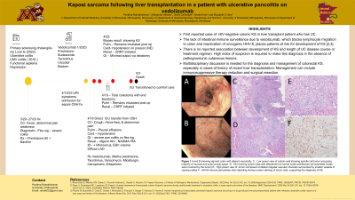
Case Description/Methods: A 28-year-old male with past medical history of primary sclerosing cholangitis status post liver transplantation (LT) on tacrolimus monotherapy, ulcerative pancolitis (UC) on vedolizumab, history of CMV related colitis, and congenital asplenia presented with fever, abdominal pain, and bloody diarrhea. A month prior, the patient was admitted for presumed UC flare and started on a prednisone taper. Initial work up revealed kidney injury, Epstein Barr viremia, edematous colon and rectum with diffuse lymphadenopathy on computed tomography (CT). Patient’s clinical status worsened to septic shock and renal failure requiring renal replacement therapy. Flexible sigmoidoscopy showed friable, inflamed mucosa with bleeding ulcers. (Figure 1a,b). No submucosal nodules were identified. Steroid therapy was escalated and prior to admission Tacrolimus and Vedolizumab continued. Given his worsening clinical course, emergent total colectomy with end ileostomy was performed. Colon histopathology samples demonstrated HHV-8-associated colonic KS with lymph node involvement (Figure 1c-f). Serum HHV-8 PCR was 3,700,000 copies/ml. HIV was undetectable. Skin examination was within normal limits. Given his ongoing critical illness, the patient pursued comfort care and died.
Discussion: This is the first report of HIV-negative HHV-8-associated colonic KS in LT recipient with UC. There is no reported association between development of KS and length of UC disease course or treatment regimen. The lack of intestinal immune surveillance due to vedolizumab, which blocks lymphocyte migration to colon and reactivation of oncogenic HHV-8, places patients at risk for development of KS. High index of suspicion is required to make this diagnosis in the absence of pathognomonic cutaneous lesions. Multidisciplinary discussion is needed for the diagnosis and management of colorectal KS, especially in cases of history of recent LT. Management can include immunosuppressive therapy reduction and surgical resection.

Disclosures:
Pavithra Ramakrishnan, MS, MD1, Mohamed A. Hassan, MD2, David Cartwright, MD2, Khalid Amin, MD3, Elizabeth S. Aby, MD2. P3670 - Kaposi Sarcoma Following Liver Transplantation in a Patient With Ulcerative Pancolitis on Vedolizumab, ACG 2023 Annual Scientific Meeting Abstracts. Vancouver, BC, Canada: American College of Gastroenterology.
Pavithra Ramakrishnan, MS, MD1, Mohamed A. Hassan, MD2, David Cartwright, MD2, Khalid Amin, MD3, Elizabeth S. Aby, MD2
1University of MInnesota, Minneapolis, MN; 2University of Minnesota, Minneapolis, MN; 3University of Minnesota Medical Center, Minneapolis, MN
Introduction: Kaposi sarcoma (KS) is a vascular tumor associated with the oncogenic virus Human Herpes Virus 8 (HHV-8). The iatrogenic variant is increasingly reported in the setting of prolonged immunosuppression, such as inflammatory bowel disease (IBD) or kidney transplant. This case highlights the importance of a broad differential diagnosis for immunosuppressed patients with IBD who are unresponsive to conventional therapy.
Case Description/Methods: A 28-year-old male with past medical history of primary sclerosing cholangitis status post liver transplantation (LT) on tacrolimus monotherapy, ulcerative pancolitis (UC) on vedolizumab, history of CMV related colitis, and congenital asplenia presented with fever, abdominal pain, and bloody diarrhea. A month prior, the patient was admitted for presumed UC flare and started on a prednisone taper. Initial work up revealed kidney injury, Epstein Barr viremia, edematous colon and rectum with diffuse lymphadenopathy on computed tomography (CT). Patient’s clinical status worsened to septic shock and renal failure requiring renal replacement therapy. Flexible sigmoidoscopy showed friable, inflamed mucosa with bleeding ulcers. (Figure 1a,b). No submucosal nodules were identified. Steroid therapy was escalated and prior to admission Tacrolimus and Vedolizumab continued. Given his worsening clinical course, emergent total colectomy with end ileostomy was performed. Colon histopathology samples demonstrated HHV-8-associated colonic KS with lymph node involvement (Figure 1c-f). Serum HHV-8 PCR was 3,700,000 copies/ml. HIV was undetectable. Skin examination was within normal limits. Given his ongoing critical illness, the patient pursued comfort care and died.
Discussion: This is the first report of HIV-negative HHV-8-associated colonic KS in LT recipient with UC. There is no reported association between development of KS and length of UC disease course or treatment regimen. The lack of intestinal immune surveillance due to vedolizumab, which blocks lymphocyte migration to colon and reactivation of oncogenic HHV-8, places patients at risk for development of KS. High index of suspicion is required to make this diagnosis in the absence of pathognomonic cutaneous lesions. Multidisciplinary discussion is needed for the diagnosis and management of colorectal KS, especially in cases of history of recent LT. Management can include immunosuppressive therapy reduction and surgical resection.

Figure: Gross and histopathologic evidence of KS Figure 1- A,B Images of sigmoid colon showing altered vascularity, congestion (edema), erythema and friability. C: Low power view of colonic wall showing spindle cell tumor occupying majority of mucosa and submucosal space. D: KS involving lymph node with effacement of normal nodal architecture (all submitted nodes were involved by the tumor) E: High power view of tumor composed of dilated irregular vascular channels surrounded by smaller vessels of varying caliber. F: HHV8 immunoperoxidase stain depicting strong nuclear staining of tumor cells, supporting the diagnosis of KS.
Disclosures:
Pavithra Ramakrishnan indicated no relevant financial relationships.
Mohamed Hassan indicated no relevant financial relationships.
David Cartwright indicated no relevant financial relationships.
Khalid Amin indicated no relevant financial relationships.
Elizabeth Aby indicated no relevant financial relationships.
Pavithra Ramakrishnan, MS, MD1, Mohamed A. Hassan, MD2, David Cartwright, MD2, Khalid Amin, MD3, Elizabeth S. Aby, MD2. P3670 - Kaposi Sarcoma Following Liver Transplantation in a Patient With Ulcerative Pancolitis on Vedolizumab, ACG 2023 Annual Scientific Meeting Abstracts. Vancouver, BC, Canada: American College of Gastroenterology.

