Tuesday Poster Session
Category: Interventional Endoscopy
P3726 - Intra-Operative Transenteric ERCP: Management of Bile Duct and Pancreatic Duct Disruption After Gunshot Injury
Tuesday, October 24, 2023
10:30 AM - 4:00 PM PT
Location: Exhibit Hall

Has Audio
- AL
Andy Lin, MD
University of California Irvine
Orange, California
Presenting Author(s)
Andy Lin, MD1, David L. Cheung, MD2, Amirali Tavangar, MD1, Khaldoon Khirfan, MD1, Joshua Kwon, MD1, Jason Samarasena, MD1, John Lee, MD1
1University of California Irvine, Orange, CA; 2UC Irvine, Orange, CA
Introduction: Intraoperative endoscopic retrograde cholangiopancreatography (IO-ERCP) is sometimes utilized for patients with altered anatomy such as Roux-en-Y gastric bypass via a laparoscopic-assisted transgastric approach. Here we present a case of a gunshot wound with common bile duct (CBD) and pancreatic duct (PD) injury treated with stenting via open transenteric IO-ERCP.
Case Description/Methods: A 19-year-old male presents after gunshot wound to the epigastrium. Emergency exploratory laparotomy revealed the gunshot wound traversing from the distal anterior stomach through to the posterior wall of the duodenum resulting in laceration of the liver, pancreatic head, as well as significant hematoma and distal gastric perforation with spillage of the gastric contents in the peritoneum. No clear bile seen but there was high suspicion about bile and pancreatic duct injury.
The stomach was stapled proximal to the antrum and gastrojejunal anastomosis was performed. Due to concern about pancreatic and bile duct injury which would affect the surgical planning, IO-ERCP was requested to perform cholangiogram and pancreatogram with possible stenting if duct injury was detected. Access to the papilla was lost due to distal gastrectomy and the newly formed anastomosis. Endoscopic ultrasound (EUS) was performed with the PD caliber about 1 mm; CBD was poorly visualized from the transgastric route. EUS-guided pancreatogram using 22-G FNA needle was limited due to the thin duct caliber and no obvious leaks were detected.
The decision was to perform an enterostomy via the distal duodenum to gain access to the papilla. Billroth 2 sphincterotome was used for cannulation. The Major and minor papilla appeared unremarkable, however pancreatogram showed extravasation near the neck. Pancreatic sphincterotomy was performed and 5 French x9cm single pigtail stent was placed. Then cholangiogram showed leak near hilum for which a 7 French x12 cm single pigtail stent was placed into the right hepatic duct. The CBD was noted to be over 75% disrupted and the stent was visible, so hepaticojejunostomy was pursued. The postoperative course was uneventful and patient was discharged.
Discussion: Peroral IO-ERCP has been reported to help guide surgical management after gunshot wounds to evaluate disruption of the pancreatic or biliary duct. In this case, we report IO-ERCP through enterostomy due to an inaccessible papilla, which was highly valuable in the detection and management of patient's bile leak.

Disclosures:
Andy Lin, MD1, David L. Cheung, MD2, Amirali Tavangar, MD1, Khaldoon Khirfan, MD1, Joshua Kwon, MD1, Jason Samarasena, MD1, John Lee, MD1. P3726 - Intra-Operative Transenteric ERCP: Management of Bile Duct and Pancreatic Duct Disruption After Gunshot Injury, ACG 2023 Annual Scientific Meeting Abstracts. Vancouver, BC, Canada: American College of Gastroenterology.
1University of California Irvine, Orange, CA; 2UC Irvine, Orange, CA
Introduction: Intraoperative endoscopic retrograde cholangiopancreatography (IO-ERCP) is sometimes utilized for patients with altered anatomy such as Roux-en-Y gastric bypass via a laparoscopic-assisted transgastric approach. Here we present a case of a gunshot wound with common bile duct (CBD) and pancreatic duct (PD) injury treated with stenting via open transenteric IO-ERCP.
Case Description/Methods: A 19-year-old male presents after gunshot wound to the epigastrium. Emergency exploratory laparotomy revealed the gunshot wound traversing from the distal anterior stomach through to the posterior wall of the duodenum resulting in laceration of the liver, pancreatic head, as well as significant hematoma and distal gastric perforation with spillage of the gastric contents in the peritoneum. No clear bile seen but there was high suspicion about bile and pancreatic duct injury.
The stomach was stapled proximal to the antrum and gastrojejunal anastomosis was performed. Due to concern about pancreatic and bile duct injury which would affect the surgical planning, IO-ERCP was requested to perform cholangiogram and pancreatogram with possible stenting if duct injury was detected. Access to the papilla was lost due to distal gastrectomy and the newly formed anastomosis. Endoscopic ultrasound (EUS) was performed with the PD caliber about 1 mm; CBD was poorly visualized from the transgastric route. EUS-guided pancreatogram using 22-G FNA needle was limited due to the thin duct caliber and no obvious leaks were detected.
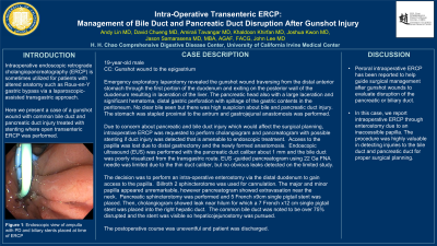
The decision was to perform an enterostomy via the distal duodenum to gain access to the papilla. Billroth 2 sphincterotome was used for cannulation. The Major and minor papilla appeared unremarkable, however pancreatogram showed extravasation near the neck. Pancreatic sphincterotomy was performed and 5 French x9cm single pigtail stent was placed. Then cholangiogram showed leak near hilum for which a 7 French x12 cm single pigtail stent was placed into the right hepatic duct. The CBD was noted to be over 75% disrupted and the stent was visible, so hepaticojejunostomy was pursued. The postoperative course was uneventful and patient was discharged.
Discussion: Peroral IO-ERCP has been reported to help guide surgical management after gunshot wounds to evaluate disruption of the pancreatic or biliary duct. In this case, we report IO-ERCP through enterostomy due to an inaccessible papilla, which was highly valuable in the detection and management of patient's bile leak.

Figure: Endoscopic view of ampulla with PD and biliary stents placed at time of ERCP
Disclosures:
Andy Lin indicated no relevant financial relationships.
David Cheung indicated no relevant financial relationships.
Amirali Tavangar indicated no relevant financial relationships.
Khaldoon Khirfan indicated no relevant financial relationships.
Joshua Kwon indicated no relevant financial relationships.
Jason Samarasena: Applied Medical – Advisor or Review Panel Member. Boston Scientific – Consultant. Conmed – Consultant. Cook – Educational Grant. Neptune Medical – Consultant. Olympus – Consultant. Steris – Consultant.
John Lee indicated no relevant financial relationships.
Andy Lin, MD1, David L. Cheung, MD2, Amirali Tavangar, MD1, Khaldoon Khirfan, MD1, Joshua Kwon, MD1, Jason Samarasena, MD1, John Lee, MD1. P3726 - Intra-Operative Transenteric ERCP: Management of Bile Duct and Pancreatic Duct Disruption After Gunshot Injury, ACG 2023 Annual Scientific Meeting Abstracts. Vancouver, BC, Canada: American College of Gastroenterology.
