Tuesday Poster Session
Category: Interventional Endoscopy
P3730 - Unveiling the Silent Intruder: A Large Gastric Schwannoma Masquerading as Worsening Dyspepsia
Tuesday, October 24, 2023
10:30 AM - 4:00 PM PT
Location: Exhibit Hall

Has Audio

Niloy Ghosh, MD
University of New Mexico Health Sciences Center
Albuquerque, NM
Presenting Author(s)
Niloy Ghosh, MD, Aamer Abbass, MD, Gulshan Parasher, MD
University of New Mexico Health Sciences Center, Albuquerque, NM
Introduction: Gastric schwannomas are rare mesenchymal neoplasms originating from Schwann cells of the peripheral nervous system. Despite their low incidence (0.2% of all gastric tumors), understanding the pathogenesis, accurate diagnosis, and appropriate management of gastric schwannomas is of significant clinical importance. In this case, we describe a potentially pre-cancerous gastric schwannoma presenting with nonspecific gastrointestinal symptoms.
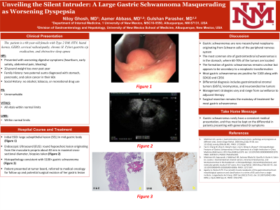
Case Description/Methods: A 68-year-old female with past medical history Type 2 DM, hypertension, hiatal hernia, GERD, cervical radiculopathy, chronic H. Pylori gastritis s/p eradication and obstructive sleep apnea presented to the emergency department for worsening digestive symptoms. The patient noted symptoms of heartburn, early satiety, upper abdominal pain and bloating in addition to 20-pound weight loss over the span of one year. Family history was significant for two paternal aunts diagnosed with stomach, pancreatic, and colon cancer in their 60s. She denied any use of tobacco, drugs, and alcohol. Initial EGD with regular adult gastroscope was unremarkable except for a large subepithelial lesion (SEL) in mid gastric body and the patient was subsequently referred for upper EUS to evaluate the SEL in the stomach. EUS revealed a round hypoechoic lesion originating from the muscularis propria about 40 mm in maximal cross-sectional diameter with fine needle biopsy with 19G needle. Histopathology revealed S100+ gastric schwannoma. Follow up was arranged with oncology, and the patient is due to return to clinic for follow up and potential surgical excision of her gastric lesion.
Discussion: Gastric schwannomas remain a rare entity, as most schwannomas are observed in the head and neck. However, the most common site of gastrointestinal schwannomas is in the stomach, where 60-70% of the tumors are located. The formation of gastric schwannomas remains unclear but appears to be secondary to a neoplastic transformation, and most gastric schwannomas are positive for S100 along with SOX10 and CD56. The differential diagnosis is broad, but includes gastrointestinal stromal tumors (GISTs), leiomyomas, and neuroendocrine tumors. As specific clinical manifestation and imaging findings of gastric schwannomas are rarely observed, management strategies vary and range from surveillance to adjuvant therapy. However, surgical resection remains the mainstay of treatment for most gastric schwannomas.

Disclosures:
Niloy Ghosh, MD, Aamer Abbass, MD, Gulshan Parasher, MD. P3730 - Unveiling the Silent Intruder: A Large Gastric Schwannoma Masquerading as Worsening Dyspepsia, ACG 2023 Annual Scientific Meeting Abstracts. Vancouver, BC, Canada: American College of Gastroenterology.
University of New Mexico Health Sciences Center, Albuquerque, NM
Introduction: Gastric schwannomas are rare mesenchymal neoplasms originating from Schwann cells of the peripheral nervous system. Despite their low incidence (0.2% of all gastric tumors), understanding the pathogenesis, accurate diagnosis, and appropriate management of gastric schwannomas is of significant clinical importance. In this case, we describe a potentially pre-cancerous gastric schwannoma presenting with nonspecific gastrointestinal symptoms.
Case Description/Methods: A 68-year-old female with past medical history Type 2 DM, hypertension, hiatal hernia, GERD, cervical radiculopathy, chronic H. Pylori gastritis s/p eradication and obstructive sleep apnea presented to the emergency department for worsening digestive symptoms. The patient noted symptoms of heartburn, early satiety, upper abdominal pain and bloating in addition to 20-pound weight loss over the span of one year. Family history was significant for two paternal aunts diagnosed with stomach, pancreatic, and colon cancer in their 60s. She denied any use of tobacco, drugs, and alcohol. Initial EGD with regular adult gastroscope was unremarkable except for a large subepithelial lesion (SEL) in mid gastric body and the patient was subsequently referred for upper EUS to evaluate the SEL in the stomach. EUS revealed a round hypoechoic lesion originating from the muscularis propria about 40 mm in maximal cross-sectional diameter with fine needle biopsy with 19G needle. Histopathology revealed S100+ gastric schwannoma. Follow up was arranged with oncology, and the patient is due to return to clinic for follow up and potential surgical excision of her gastric lesion.
Discussion: Gastric schwannomas remain a rare entity, as most schwannomas are observed in the head and neck. However, the most common site of gastrointestinal schwannomas is in the stomach, where 60-70% of the tumors are located. The formation of gastric schwannomas remains unclear but appears to be secondary to a neoplastic transformation, and most gastric schwannomas are positive for S100 along with SOX10 and CD56. The differential diagnosis is broad, but includes gastrointestinal stromal tumors (GISTs), leiomyomas, and neuroendocrine tumors. As specific clinical manifestation and imaging findings of gastric schwannomas are rarely observed, management strategies vary and range from surveillance to adjuvant therapy. However, surgical resection remains the mainstay of treatment for most gastric schwannomas.

Figure: Subepithelial Lesion in Gastric Body
Disclosures:
Niloy Ghosh indicated no relevant financial relationships.
Aamer Abbass indicated no relevant financial relationships.
Gulshan Parasher indicated no relevant financial relationships.
Niloy Ghosh, MD, Aamer Abbass, MD, Gulshan Parasher, MD. P3730 - Unveiling the Silent Intruder: A Large Gastric Schwannoma Masquerading as Worsening Dyspepsia, ACG 2023 Annual Scientific Meeting Abstracts. Vancouver, BC, Canada: American College of Gastroenterology.
