Tuesday Poster Session
Category: Interventional Endoscopy
P3731 - Nonsurgical Management of Mirizzi Syndrome With Cholecystoscopy and Electrohydraulic Lithotripsy
Tuesday, October 24, 2023
10:30 AM - 4:00 PM PT
Location: Exhibit Hall

Has Audio
- CE
Carter Edmunds, MD
University of Alabama at Birmingham Hospital
Birmingham, AL
Presenting Author(s)
Carter Edmunds, MD1, Swati Pawa, MD2
1University of Alabama at Birmingham Hospital, Birmingham, AL; 2Atrium Health Wake Forest Baptist, Advance, NC
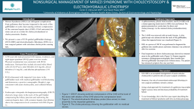
Introduction: Mirizzi Syndrome (MS) is an infrequent complication from gallstones that become impacted in the neck of the gallbladder or cystic duct causing extrinsic compression of the common hepatic duct (CHD). If left untreated, the stone can act as a nidus for cholecystoduodenal or cholecystoenteric fistulas. We present a case of EUS-guided gallbladder drainage (EUS-GBD) and electrohydraulic lithotripsy (EHL) in a non-surgical patient with calculous cholecystitis causing MS.
Case Description/Methods: A 94-year-old male presented with nausea, weakness, and right upper quadrant (RUQ) pain over two weeks. Physical examination was consistent with RUQ tenderness. Pertinent labs include elevated white blood cells (24.4x10³/µL), total bilirubin (4.8 mg/dL), direct bilirubin (3.4 mg/ld.), and alkaline phosphatase (154 U/L). RUQ ultrasound demonstrated an impacted 2cm stone in the gallbladder neck with marked gallbladder wall thickening and distension, a positive sonographic Murphy's sign consistent with acute cholecystitis, and a dilated biliary tree. Endoscopic retrograde cholangiopancreatography (ERCP) showed a large stone in the neck of the gall bladder compressing the biliary junction and narrowing the common hepatic duct, with common hepatic duct dilation above the compression level consistent with MS (Figure 1). A sphincterotomy, plastic stent insertion, and EUS-GBD with a lumen apposing metal stent (LAMS) were performed. The patient presented six weeks later for direct oral cholecystoscopy via the matured cholecystoduodenostomy tract. The LAMS was removed with rat tooth forceps. A slim gastroscope was advanced into the neck of the gallbladder, where the impacted stone was visualized (Figure 2). EHL at a power of 100 W was performed to fragment the gallstone into smaller pieces and stone clearance was achieved after two sessions. Final inspection on direct cholecystoscopy showed no retained stone in the gallbladder neck (Figure 3). Repeat magnetic resonance cholangiopancreatography three months later showed gallbladder decompression without stones. The patient has remained symptom free one year later.
Discussion: EUS-GBD is an accepted management of acute calculus cholecystitis in patients who are poor surgical candidates. However, the role of concomitant endoscopic lithotripsy in non-surgical candidates remains unclear. To our knowledge, this is the first case report describing the treatment of MS with EHL via cholecystoscopy.

Disclosures:
Carter Edmunds, MD1, Swati Pawa, MD2. P3731 - Nonsurgical Management of Mirizzi Syndrome With Cholecystoscopy and Electrohydraulic Lithotripsy, ACG 2023 Annual Scientific Meeting Abstracts. Vancouver, BC, Canada: American College of Gastroenterology.
1University of Alabama at Birmingham Hospital, Birmingham, AL; 2Atrium Health Wake Forest Baptist, Advance, NC
Introduction: Mirizzi Syndrome (MS) is an infrequent complication from gallstones that become impacted in the neck of the gallbladder or cystic duct causing extrinsic compression of the common hepatic duct (CHD). If left untreated, the stone can act as a nidus for cholecystoduodenal or cholecystoenteric fistulas. We present a case of EUS-guided gallbladder drainage (EUS-GBD) and electrohydraulic lithotripsy (EHL) in a non-surgical patient with calculous cholecystitis causing MS.
Case Description/Methods: A 94-year-old male presented with nausea, weakness, and right upper quadrant (RUQ) pain over two weeks. Physical examination was consistent with RUQ tenderness. Pertinent labs include elevated white blood cells (24.4x10³/µL), total bilirubin (4.8 mg/dL), direct bilirubin (3.4 mg/ld.), and alkaline phosphatase (154 U/L). RUQ ultrasound demonstrated an impacted 2cm stone in the gallbladder neck with marked gallbladder wall thickening and distension, a positive sonographic Murphy's sign consistent with acute cholecystitis, and a dilated biliary tree. Endoscopic retrograde cholangiopancreatography (ERCP) showed a large stone in the neck of the gall bladder compressing the biliary junction and narrowing the common hepatic duct, with common hepatic duct dilation above the compression level consistent with MS (Figure 1). A sphincterotomy, plastic stent insertion, and EUS-GBD with a lumen apposing metal stent (LAMS) were performed. The patient presented six weeks later for direct oral cholecystoscopy via the matured cholecystoduodenostomy tract. The LAMS was removed with rat tooth forceps. A slim gastroscope was advanced into the neck of the gallbladder, where the impacted stone was visualized (Figure 2). EHL at a power of 100 W was performed to fragment the gallstone into smaller pieces and stone clearance was achieved after two sessions. Final inspection on direct cholecystoscopy showed no retained stone in the gallbladder neck (Figure 3). Repeat magnetic resonance cholangiopancreatography three months later showed gallbladder decompression without stones. The patient has remained symptom free one year later.
Discussion: EUS-GBD is an accepted management of acute calculus cholecystitis in patients who are poor surgical candidates. However, the role of concomitant endoscopic lithotripsy in non-surgical candidates remains unclear. To our knowledge, this is the first case report describing the treatment of MS with EHL via cholecystoscopy.

Figure: Figure 1: ERCP showing extrinsic compression of the CHD at the level of the stone with dilation of the CHD above the compression level. Figure 2: The electrohydraulic lithotripsy probe (blue arrow) in close proximity to the impacted gallstone. Figure 3: The cholecystoscopy showing the gallbladder with no residual stones.
Disclosures:
Carter Edmunds indicated no relevant financial relationships.
Swati Pawa: boston scientific corporation – Consultant.
Carter Edmunds, MD1, Swati Pawa, MD2. P3731 - Nonsurgical Management of Mirizzi Syndrome With Cholecystoscopy and Electrohydraulic Lithotripsy, ACG 2023 Annual Scientific Meeting Abstracts. Vancouver, BC, Canada: American College of Gastroenterology.

