Tuesday Poster Session
Category: Interventional Endoscopy
P3756 - Endoscopic Embolization of Large Duodenal Varices Under Fluoroscopy Guidance
Tuesday, October 24, 2023
10:30 AM - 4:00 PM PT
Location: Exhibit Hall

Has Audio

Mahinaz Mohsen, BA
Rutgers New Jersey Medical School
Newark, NJ
Presenting Author(s)
Mahinaz Mohsen, BA, Yazan Abboud, MD, Kaveh Hajifathalian, MD, MPH
Rutgers New Jersey Medical School, Newark, NJ
Introduction: Ectopic varices are those found outside the gastroesophageal region. Bleeding ectopic varices account for 2-5% of total variceal bleeding. While rare, ectopic variceal bleeding can lead to severe hemorrhage with a mortality rate of 40%. Bleeding duodenal varices are even rarer, making up 17% of total bleeding ectopic varices. We present a case of large, bleeding duodenal varices that were managed endoscopically.
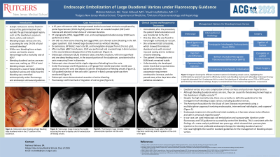
Case Description/Methods: A 67-year-old woman with decompensated alcohol-associated cirrhosis complicated by portal hypertension (Child-Pugh B) presented from an outside hospital (OSH) with melena and confusion. Blood pressure was 96/62, heart rate 99, and hemoglobin 8.2 g/dL. CT angiography (CTA) and tagged RBC scan were performed at OSH. CTA did not show active bleeding but tagged RBC scan showed active bleeding in the left upper quadrant. Esophagogastroduodenoscopy (EGD) was performed and revealed large ( >5mm) varices in the second portion of the duodenum (Figure 1). Endoscopic ultrasound showed a tubal, anechoic structure, with one superficial and one deep feeding vessel, in the second portion of the duodenum, consistent with a varix measuring 8 mm in diameter. Endoscopic view showed white nipple stigmata of bleeding. Under fluoroscopy and EUS guidance, a 19-gauge fine-needle-aspiration needle was used to access the varix and deploy 4 coils for embolization of feeding vessels (Figure 2). EUS-guided injection of the varix with 1 gram of n-Butyl cyanoacrylate was then conducted (Figure 3). Endoscopic exam demonstrated cessation of active bleeding and fluoroscopy confirmed lack of migration of coil or glue (Figure 4). Immediately after the procedure, patient failed extubation and was transferred to the ICU. One day later, she was found to have bright red blood per rectum. Repeat EGD showed thrombosed duodenal varix with minimal mucosal oozing but no evidence of active bleeding. Over the course of the following week, the patient developed sepsis and her vasopressor requirement continued to increase. She was transitioned to hospice care and passed away after palliative extubation.
Discussion: There are currently no definitive guidelines for the management of bleeding ectopic varices. Management options include endoscopic band ligation or sclerotherapy, transjugular intrahepatic portosystemic shunt, and surgery. We present a case of duodenal variceal bleeding that was successfully controlled via endoscopic coil embolization and cyanoacrylate injection under fluoroscopy and EUS guidance.

Disclosures:
Mahinaz Mohsen, BA, Yazan Abboud, MD, Kaveh Hajifathalian, MD, MPH. P3756 - Endoscopic Embolization of Large Duodenal Varices Under Fluoroscopy Guidance, ACG 2023 Annual Scientific Meeting Abstracts. Vancouver, BC, Canada: American College of Gastroenterology.
Rutgers New Jersey Medical School, Newark, NJ
Introduction: Ectopic varices are those found outside the gastroesophageal region. Bleeding ectopic varices account for 2-5% of total variceal bleeding. While rare, ectopic variceal bleeding can lead to severe hemorrhage with a mortality rate of 40%. Bleeding duodenal varices are even rarer, making up 17% of total bleeding ectopic varices. We present a case of large, bleeding duodenal varices that were managed endoscopically.
Case Description/Methods: A 67-year-old woman with decompensated alcohol-associated cirrhosis complicated by portal hypertension (Child-Pugh B) presented from an outside hospital (OSH) with melena and confusion. Blood pressure was 96/62, heart rate 99, and hemoglobin 8.2 g/dL. CT angiography (CTA) and tagged RBC scan were performed at OSH. CTA did not show active bleeding but tagged RBC scan showed active bleeding in the left upper quadrant. Esophagogastroduodenoscopy (EGD) was performed and revealed large ( >5mm) varices in the second portion of the duodenum (Figure 1). Endoscopic ultrasound showed a tubal, anechoic structure, with one superficial and one deep feeding vessel, in the second portion of the duodenum, consistent with a varix measuring 8 mm in diameter. Endoscopic view showed white nipple stigmata of bleeding. Under fluoroscopy and EUS guidance, a 19-gauge fine-needle-aspiration needle was used to access the varix and deploy 4 coils for embolization of feeding vessels (Figure 2). EUS-guided injection of the varix with 1 gram of n-Butyl cyanoacrylate was then conducted (Figure 3). Endoscopic exam demonstrated cessation of active bleeding and fluoroscopy confirmed lack of migration of coil or glue (Figure 4). Immediately after the procedure, patient failed extubation and was transferred to the ICU. One day later, she was found to have bright red blood per rectum. Repeat EGD showed thrombosed duodenal varix with minimal mucosal oozing but no evidence of active bleeding. Over the course of the following week, the patient developed sepsis and her vasopressor requirement continued to increase. She was transitioned to hospice care and passed away after palliative extubation.
Discussion: There are currently no definitive guidelines for the management of bleeding ectopic varices. Management options include endoscopic band ligation or sclerotherapy, transjugular intrahepatic portosystemic shunt, and surgery. We present a case of duodenal variceal bleeding that was successfully controlled via endoscopic coil embolization and cyanoacrylate injection under fluoroscopy and EUS guidance.

Figure: Figure 1 shows endoscopic view of large (>5mm) varices in the second portion of the duodenum. Figure 2 shows a 19-gauge fine-needle-aspiration needle deploying coils into the varix. Figure 3 shows injection of the varix with cyanoacrylate. Figure 4 shows lack of migration of coil or glue after coil embolization and cyanoacrylate injection were completed.
Disclosures:
Mahinaz Mohsen indicated no relevant financial relationships.
Yazan Abboud indicated no relevant financial relationships.
Kaveh Hajifathalian indicated no relevant financial relationships.
Mahinaz Mohsen, BA, Yazan Abboud, MD, Kaveh Hajifathalian, MD, MPH. P3756 - Endoscopic Embolization of Large Duodenal Varices Under Fluoroscopy Guidance, ACG 2023 Annual Scientific Meeting Abstracts. Vancouver, BC, Canada: American College of Gastroenterology.
