Tuesday Poster Session
Category: Interventional Endoscopy
P3740 - Perisplenic Hematoma as a Rare Complication of ERCP: A Case Report
Tuesday, October 24, 2023
10:30 AM - 4:00 PM PT
Location: Exhibit Hall

Has Audio

Tahne Vongsavath, DO
Kirk Kerkorian School of Medicine
Las Vegas, NV
Presenting Author(s)
Tahne Vongsavath, DO1, Nader Mehregani, 1, Kyaw Min Tun, DO2
1Kirk Kerkorian School of Medicine, Las Vegas, NV; 2Kirk Kerkorian School of Medicine at UNLV, Las Vegas, NV
Introduction: Splenic injury after endoscopy is a rare complication with less than 25 reported cases, the majority of which following colonoscopy. Endoscopic retrograde cholangiopancreatography (ERCP) is a rare etiology for splenic injury. This case reports on a patient who developed perisplenic hematoma after undergoing ERCP to receive metallic common bile duct stent for obstructive pancreatic head mass.
Case Description/Methods: A 57-year-old female with history of non-alcoholic fatty liver presented to the hospital with 7 months of abdominal pain and jaundice with acholic stools. Pancreatitis was excluded as lipase was within reference level and abdominal imaging showed no pancreatitis. CT abdomen/pelvis was concerning for diffuse metastatic lesions. The following labs were elevated: ALT 436, AST 407, alkaline phosphatase 403, total bilirubin 15, GGT 493, CA 19-9 6888. ERCP with sphincterotomy was performed. A metal stent was placed at the common bile duct and fine needle aspiration of one the pancreatic head lesions showed adenocarcinoma. No complications were noted post-ERCP based on clinical examination and laboratory profile. The following day, CT chest imaging was done to further evaluate metastatic lesions and was found to have acute perisplenic hematoma at the inferior margin of left hemidiaphragm with an area of acute extravasation. The patient was evaluated and found in stable condition. It was new compared to imaging the day prior, and the patient was without any new trauma or inciting events other than ERCP. The patient remained hemodynamically stable without sequelae of acute blood loss. The patient was treated conservatively and did not receive repeat imaging.
Discussion: Splenic injury following ERCP is an uncommon life-threatening complication. There must be a high index of suspicion in any patient who develops abdominal pain, anemia, or hypotension after ERCP. This patient did not have indication of bleeding until a repeat CT uncovered a perisplenic hematoma. Although the etiology of ERCP-related splenic injury is not fully known, it may result from excessive traction. Predisposing factors may include intra-abdominal adhesions from previous surgery, chronic pancreatitis, prolonged procedure, stomach insufflation, or obstructing tumor. This may cause increased tension on splenic ligaments causing traction on surrounding tissue. Previous case reports noted patients with splenic lacerations and ruptures underwent splenectomies, and those with splenic hematomas were treated conservatively.

Disclosures:
Tahne Vongsavath, DO1, Nader Mehregani, 1, Kyaw Min Tun, DO2. P3740 - Perisplenic Hematoma as a Rare Complication of ERCP: A Case Report, ACG 2023 Annual Scientific Meeting Abstracts. Vancouver, BC, Canada: American College of Gastroenterology.
1Kirk Kerkorian School of Medicine, Las Vegas, NV; 2Kirk Kerkorian School of Medicine at UNLV, Las Vegas, NV
Introduction: Splenic injury after endoscopy is a rare complication with less than 25 reported cases, the majority of which following colonoscopy. Endoscopic retrograde cholangiopancreatography (ERCP) is a rare etiology for splenic injury. This case reports on a patient who developed perisplenic hematoma after undergoing ERCP to receive metallic common bile duct stent for obstructive pancreatic head mass.
Case Description/Methods: A 57-year-old female with history of non-alcoholic fatty liver presented to the hospital with 7 months of abdominal pain and jaundice with acholic stools. Pancreatitis was excluded as lipase was within reference level and abdominal imaging showed no pancreatitis. CT abdomen/pelvis was concerning for diffuse metastatic lesions. The following labs were elevated: ALT 436, AST 407, alkaline phosphatase 403, total bilirubin 15, GGT 493, CA 19-9 6888. ERCP with sphincterotomy was performed. A metal stent was placed at the common bile duct and fine needle aspiration of one the pancreatic head lesions showed adenocarcinoma. No complications were noted post-ERCP based on clinical examination and laboratory profile. The following day, CT chest imaging was done to further evaluate metastatic lesions and was found to have acute perisplenic hematoma at the inferior margin of left hemidiaphragm with an area of acute extravasation. The patient was evaluated and found in stable condition. It was new compared to imaging the day prior, and the patient was without any new trauma or inciting events other than ERCP. The patient remained hemodynamically stable without sequelae of acute blood loss. The patient was treated conservatively and did not receive repeat imaging.
Discussion: Splenic injury following ERCP is an uncommon life-threatening complication. There must be a high index of suspicion in any patient who develops abdominal pain, anemia, or hypotension after ERCP. This patient did not have indication of bleeding until a repeat CT uncovered a perisplenic hematoma. Although the etiology of ERCP-related splenic injury is not fully known, it may result from excessive traction. Predisposing factors may include intra-abdominal adhesions from previous surgery, chronic pancreatitis, prolonged procedure, stomach insufflation, or obstructing tumor. This may cause increased tension on splenic ligaments causing traction on surrounding tissue. Previous case reports noted patients with splenic lacerations and ruptures underwent splenectomies, and those with splenic hematomas were treated conservatively.

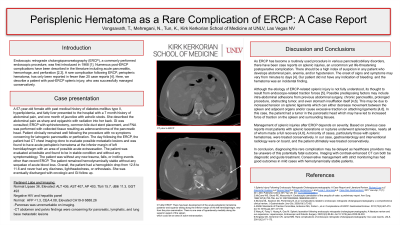
Figure: Figure 1: Initial CT scan showing the perisplenic area. Figure 2: Follow up CT scan showing developing area of perisplenic hematoma with an area of possible acute extravasation.
Disclosures:
Tahne Vongsavath indicated no relevant financial relationships.
Nader Mehregani indicated no relevant financial relationships.
Kyaw Min Tun indicated no relevant financial relationships.
Tahne Vongsavath, DO1, Nader Mehregani, 1, Kyaw Min Tun, DO2. P3740 - Perisplenic Hematoma as a Rare Complication of ERCP: A Case Report, ACG 2023 Annual Scientific Meeting Abstracts. Vancouver, BC, Canada: American College of Gastroenterology.
