Tuesday Poster Session
Category: Interventional Endoscopy
P3742 - Bouveret Syndrome as a Cause of Gastric Outlet Obstruction: A Case Report
Tuesday, October 24, 2023
10:30 AM - 4:00 PM PT
Location: Exhibit Hall

Has Audio
- SG
Santiago Garcia Ortiz, MD
Creighton University
Phoenix, AZ
Presenting Author(s)
Santiago Garcia Ortiz, MD1, Neha Khan, MD1, Kayvon Sotoudeh, MD1, Aida Rezaie, MD2, Neil Vyas, MD1, Savio Reddymasu, MD1
1Creighton University, Phoenix, AZ; 2Creighton University, Mesa, AZ
Introduction: Bouveret syndrome is a rare complication of cholelithiasis. Delay in diagnosis is associated with a high mortality rate. It is a rare form of small bowel obstruction caused by a large biliary stone entering the lumen of the duodenum or the stomach through a bilioenteric fistula. Diagnosis is based upon clinical manifestations, endoscopy, and imaging studies such as abdominal computed tomography (CT) and magnetic resonance cholangiopancreatography (MRCP). Treatment options include various surgical techniques. However, recent advances in endoscopy also allow for non-surgical endoscopic treatment options.
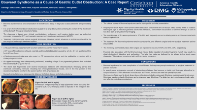
Case Description/Methods: 79 year old male presented with recurrent abdominal pain for more than 6 weeks. A CT scan of the abdomen showed a partial gastric outlet obstruction caused by a 5.5 x 3.4 cm gallstone at the level of the pylorus. A fistulous communication was also noted between the pylorus and the gallbladder consistent with a cholecystogastric fistula. On upper endoscopy, a large 5 cm pigmented gallstone was visualized that occluded the duodenal bulb. The stone was fragmented over several endoscopic sessions with electrohydraulic lithotripsy (EHL) and eventually completed removed, revealing two cholecystoduodenal fistulas, one in the first portion of the duodenum and another in the second portion. Surgical consultation was subsequently obtained for fistula closure and cholecystectomy.
Discussion: Bouveret syndrome is a rare complication of cholelithiasis that requires prompt endoscopic or surgical treatment to prevent mortality. In recent years, endoscopic removal of obstructing gallstones has become a viable, well tolerated alternative to surgical treatment.
With advances in endoscopic techniques, the success rate has greatly improved in the recent years. Common methods used to break the large stone into pieces include endoscopic lithotripsy, extracorporeal shock wave lithotripsy, electro-hydraulic lithotripsy, and endoscopic laser lithotripsy. The latter two technologies are simple, safe, and effective, with few complications, and are currently the most commonly used methods.

Disclosures:
Santiago Garcia Ortiz, MD1, Neha Khan, MD1, Kayvon Sotoudeh, MD1, Aida Rezaie, MD2, Neil Vyas, MD1, Savio Reddymasu, MD1. P3742 - Bouveret Syndrome as a Cause of Gastric Outlet Obstruction: A Case Report, ACG 2023 Annual Scientific Meeting Abstracts. Vancouver, BC, Canada: American College of Gastroenterology.
1Creighton University, Phoenix, AZ; 2Creighton University, Mesa, AZ
Introduction: Bouveret syndrome is a rare complication of cholelithiasis. Delay in diagnosis is associated with a high mortality rate. It is a rare form of small bowel obstruction caused by a large biliary stone entering the lumen of the duodenum or the stomach through a bilioenteric fistula. Diagnosis is based upon clinical manifestations, endoscopy, and imaging studies such as abdominal computed tomography (CT) and magnetic resonance cholangiopancreatography (MRCP). Treatment options include various surgical techniques. However, recent advances in endoscopy also allow for non-surgical endoscopic treatment options.
Case Description/Methods: 79 year old male presented with recurrent abdominal pain for more than 6 weeks. A CT scan of the abdomen showed a partial gastric outlet obstruction caused by a 5.5 x 3.4 cm gallstone at the level of the pylorus. A fistulous communication was also noted between the pylorus and the gallbladder consistent with a cholecystogastric fistula. On upper endoscopy, a large 5 cm pigmented gallstone was visualized that occluded the duodenal bulb. The stone was fragmented over several endoscopic sessions with electrohydraulic lithotripsy (EHL) and eventually completed removed, revealing two cholecystoduodenal fistulas, one in the first portion of the duodenum and another in the second portion. Surgical consultation was subsequently obtained for fistula closure and cholecystectomy.
Discussion: Bouveret syndrome is a rare complication of cholelithiasis that requires prompt endoscopic or surgical treatment to prevent mortality. In recent years, endoscopic removal of obstructing gallstones has become a viable, well tolerated alternative to surgical treatment.
With advances in endoscopic techniques, the success rate has greatly improved in the recent years. Common methods used to break the large stone into pieces include endoscopic lithotripsy, extracorporeal shock wave lithotripsy, electro-hydraulic lithotripsy, and endoscopic laser lithotripsy. The latter two technologies are simple, safe, and effective, with few complications, and are currently the most commonly used methods.

Figure: Figure 1A-E: Upper endoscopy shows a large 5 cm pigmented gallstone was visualized that occluded the duodenal bulb. The stone was fragmented over several endoscopic sessions with electrohydraulic lithotripsy (EHL) and removed with a Roth Net retriever.
Disclosures:
Santiago Garcia Ortiz indicated no relevant financial relationships.
Neha Khan indicated no relevant financial relationships.
Kayvon Sotoudeh indicated no relevant financial relationships.
Aida Rezaie indicated no relevant financial relationships.
Neil Vyas indicated no relevant financial relationships.
Savio Reddymasu indicated no relevant financial relationships.
Santiago Garcia Ortiz, MD1, Neha Khan, MD1, Kayvon Sotoudeh, MD1, Aida Rezaie, MD2, Neil Vyas, MD1, Savio Reddymasu, MD1. P3742 - Bouveret Syndrome as a Cause of Gastric Outlet Obstruction: A Case Report, ACG 2023 Annual Scientific Meeting Abstracts. Vancouver, BC, Canada: American College of Gastroenterology.
