Tuesday Poster Session
Category: Interventional Endoscopy
P3747 - Endoscopic Mucosal Incisional, Closure, ResectiON (E-MICRON): A Novel Technique for Safe and Efficient En-Bloc Foregut Resection
Tuesday, October 24, 2023
10:30 AM - 4:00 PM PT
Location: Exhibit Hall

Has Audio

Talia F. Malik, MD
Chicago Medical School at Rosalind Franklin University of Medicine and Science
North Chicago, IL
Presenting Author(s)
Award: Presidential Poster Award
Talia F.. Malik, MD1, Tilak Shah, MD2
1Chicago Medical School at Rosalind Franklin University of Medicine and Science, North Chicago, IL; 2Digestive Disease Institute, Cleveland Clinic Florida, Weston, FL
Introduction: Endoscopic mucosal resection (EMR) is appropriate for en bloc resection of gastric intramucosal lesions < 15 mm in size, but endoscopic submucosal dissection (ESD) is required for larger lesions. ESD is technically challenging, labor intensive and reimbursement is rarely commensurate with the time required to perform it in the US. We describe a novel technique that combines the simplest aspects of submucosal dissection with over-the-scope clip (OTSC) placement and snare cautery to achieve en bloc resection in a safe and efficient manner.
E-MICRON technique:
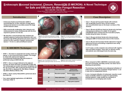
Step 1 -The lesion is inspected and cautery marks are placed 1-2 mm around the lesion margins (C). The margin is raised with submucosal injection. Circumferential Endoscopic Mucosal Incision is performed using an ESD knife (D).
Step 2 -Once no longer tethered to the adjacent mucosa, the lesion becomes considerably smaller. The endoscopist can suction the lesion completely into the cap of an OTSC. After suctioning the lesion into the cap, the OTSC is deployed (E). We utilize a Padlock clip (Steris Medical, Mentor, OH).
Step 3 -Snare cautery ResectiON is performed above the OTSC (F).
Case Description/Methods: Four cases highlight applications of E-MICRON technique(Table 1)
Case 1: 70-year-old Caucasian woman with cirrhosis was incidentally found to have a 25-mm sessile non-circumferential mass on the lesser curvature of the stomach (A).
Case 2: 68-year-old African American woman who had been undergoing surveillance of a stable 10 mm gastric subepithelial lesion arising from the muscularis propria, located along the lesser curvature (B).
Case 3: 66-year-old Chinese woman who had previously undergone EMR of intramucosal cancer at the incisura.
Case 4: 88-year-old Caucasian woman referred for irregular appearing mucosa surrounding a superficial ulcer along the lesser curvature.
In all 4 cases, en bloc resection was achieved with negative deep and lateral margins. There were no adverse events, and patients were discharged home the same day.
Discussion: When compared to ESD, E-MICRON is technically easier, less time-consuming, and likely carries a far lower risk of bleeding and perforation. The technique may facilitate more widespread utilization of endoscopic resection for neoplastic foregut lesions > 2 cm in size and for subepithelial tumors. In turn, increased utilization of endoscopic resection could reduce morbidity related to surgery for appropriately selected patients. Larger studies are necessary to corroborate our encouraging early results.

Disclosures:
Talia F.. Malik, MD1, Tilak Shah, MD2. P3747 - Endoscopic Mucosal Incisional, Closure, ResectiON (E-MICRON): A Novel Technique for Safe and Efficient En-Bloc Foregut Resection, ACG 2023 Annual Scientific Meeting Abstracts. Vancouver, BC, Canada: American College of Gastroenterology.
Talia F.. Malik, MD1, Tilak Shah, MD2
1Chicago Medical School at Rosalind Franklin University of Medicine and Science, North Chicago, IL; 2Digestive Disease Institute, Cleveland Clinic Florida, Weston, FL
Introduction: Endoscopic mucosal resection (EMR) is appropriate for en bloc resection of gastric intramucosal lesions < 15 mm in size, but endoscopic submucosal dissection (ESD) is required for larger lesions. ESD is technically challenging, labor intensive and reimbursement is rarely commensurate with the time required to perform it in the US. We describe a novel technique that combines the simplest aspects of submucosal dissection with over-the-scope clip (OTSC) placement and snare cautery to achieve en bloc resection in a safe and efficient manner.
E-MICRON technique:
Step 1 -The lesion is inspected and cautery marks are placed 1-2 mm around the lesion margins (C). The margin is raised with submucosal injection. Circumferential Endoscopic Mucosal Incision is performed using an ESD knife (D).
Step 2 -Once no longer tethered to the adjacent mucosa, the lesion becomes considerably smaller. The endoscopist can suction the lesion completely into the cap of an OTSC. After suctioning the lesion into the cap, the OTSC is deployed (E). We utilize a Padlock clip (Steris Medical, Mentor, OH).
Step 3 -Snare cautery ResectiON is performed above the OTSC (F).
Case Description/Methods: Four cases highlight applications of E-MICRON technique(Table 1)
Case 1: 70-year-old Caucasian woman with cirrhosis was incidentally found to have a 25-mm sessile non-circumferential mass on the lesser curvature of the stomach (A).
Case 2: 68-year-old African American woman who had been undergoing surveillance of a stable 10 mm gastric subepithelial lesion arising from the muscularis propria, located along the lesser curvature (B).
Case 3: 66-year-old Chinese woman who had previously undergone EMR of intramucosal cancer at the incisura.
Case 4: 88-year-old Caucasian woman referred for irregular appearing mucosa surrounding a superficial ulcer along the lesser curvature.
In all 4 cases, en bloc resection was achieved with negative deep and lateral margins. There were no adverse events, and patients were discharged home the same day.
Discussion: When compared to ESD, E-MICRON is technically easier, less time-consuming, and likely carries a far lower risk of bleeding and perforation. The technique may facilitate more widespread utilization of endoscopic resection for neoplastic foregut lesions > 2 cm in size and for subepithelial tumors. In turn, increased utilization of endoscopic resection could reduce morbidity related to surgery for appropriately selected patients. Larger studies are necessary to corroborate our encouraging early results.

Figure: Figure 1. Endoscopic Mucosal Incisional Closure Resection (E-MICRON) technique for en bloc foregut resection.
Disclosures:
Talia Malik indicated no relevant financial relationships.
Tilak Shah: Steris Medical – Consultant.
Talia F.. Malik, MD1, Tilak Shah, MD2. P3747 - Endoscopic Mucosal Incisional, Closure, ResectiON (E-MICRON): A Novel Technique for Safe and Efficient En-Bloc Foregut Resection, ACG 2023 Annual Scientific Meeting Abstracts. Vancouver, BC, Canada: American College of Gastroenterology.

