Tuesday Poster Session
Category: GI Bleeding
P3508 - When it Bleeds, it Pours: Esophageal Dieulafoy as a Rare Cause of Recurrent GI Bleeding
Tuesday, October 24, 2023
10:30 AM - 4:00 PM PT
Location: Exhibit Hall

- JC
Jacqueline N. Chu, MD
Integrated Gastroenterology Consultants
North Chelmsford, MA
Presenting Author(s)
Jacqueline N.. Chu, MD
Integrated Gastroenterology Consultants, North Chelmsford, MA
Introduction: Dieulafoy lesions are a well-known cause of recurrent overt obscure gastrointestinal bleeding. These cases can be frustrating due to the transient nature of the bleed and difficulty finding the source, resulting in frequent negative endoscopic evaluations. Though Dieulafoys are classically located in the proximal stomach, esophageal Dieulafoys have been rarely reported and may be the culprit lesion in some cases with otherwise “negative” upper endoscopies.
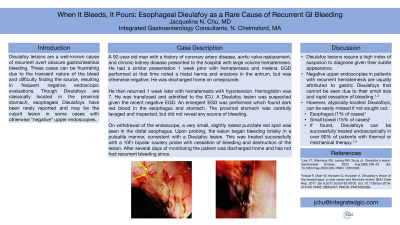
Case Description/Methods: A 92 year old man with a history of coronary artery disease, aortic valve replacement, and chronic kidney disease presented to the hospital with large volume hematemesis. He had a similar presentation 1 week prior with hematemesis and melena. EGD performed at that time noted a hiatal hernia and erosions in the antrum, but was otherwise negative. He was discharged home on omeprazole. He then returned 1 week later with hematemesis with hypotension. Hemoglobin was 7. He was transfused and admitted to the ICU. A Dieulafoy lesion was suspected given the recent negative EGD. An emergent EGD was performed which found dark red blood in the esophagus and stomach. The proximal stomach was carefully lavaged and inspected, but did not reveal any source of bleeding. On withdrawal of the endoscope, a very small, slightly raised punctate red spot was seen in the distal esophagus. Upon probing, the lesion began bleeding briskly in a pulsatile manner, consistent with a Dieulafoy lesion. This was treated successfully with a 10Fr bipolar cautery probe with cessation of bleeding and destruction of the lesion. After several days of monitoring the patient was discharged home and has not had recurrent bleeding since.
Discussion: Dieulafoy lesions require a high index of suspicion to diagnose given their subtle appearance. Negative upper endoscopies in patients with recurrent hematemesis are usually attributed to gastric Dieulafoys that cannot be seen due to their small size and rapid cessation of bleeding. However, atypically-located Dieulafoys, such as those in the esophagus, can be easily missed if not sought out. This case highlights the need for careful evaluation for small lesions throughout the GI tract that could represent a Dieulafoy, not only in the stomach, but also in the esophagus (1% of cases) and small bowel (15% of cases). If found, Dieulafoys can be successfully treated endoscopically in over 90% of patients with thermal or mechanical therapy.
Disclosures:
Jacqueline N.. Chu, MD. P3508 - When it Bleeds, it Pours: Esophageal Dieulafoy as a Rare Cause of Recurrent GI Bleeding, ACG 2023 Annual Scientific Meeting Abstracts. Vancouver, BC, Canada: American College of Gastroenterology.
Integrated Gastroenterology Consultants, North Chelmsford, MA
Introduction: Dieulafoy lesions are a well-known cause of recurrent overt obscure gastrointestinal bleeding. These cases can be frustrating due to the transient nature of the bleed and difficulty finding the source, resulting in frequent negative endoscopic evaluations. Though Dieulafoys are classically located in the proximal stomach, esophageal Dieulafoys have been rarely reported and may be the culprit lesion in some cases with otherwise “negative” upper endoscopies.
Case Description/Methods: A 92 year old man with a history of coronary artery disease, aortic valve replacement, and chronic kidney disease presented to the hospital with large volume hematemesis. He had a similar presentation 1 week prior with hematemesis and melena. EGD performed at that time noted a hiatal hernia and erosions in the antrum, but was otherwise negative. He was discharged home on omeprazole. He then returned 1 week later with hematemesis with hypotension. Hemoglobin was 7. He was transfused and admitted to the ICU. A Dieulafoy lesion was suspected given the recent negative EGD. An emergent EGD was performed which found dark red blood in the esophagus and stomach. The proximal stomach was carefully lavaged and inspected, but did not reveal any source of bleeding. On withdrawal of the endoscope, a very small, slightly raised punctate red spot was seen in the distal esophagus. Upon probing, the lesion began bleeding briskly in a pulsatile manner, consistent with a Dieulafoy lesion. This was treated successfully with a 10Fr bipolar cautery probe with cessation of bleeding and destruction of the lesion. After several days of monitoring the patient was discharged home and has not had recurrent bleeding since.
Discussion: Dieulafoy lesions require a high index of suspicion to diagnose given their subtle appearance. Negative upper endoscopies in patients with recurrent hematemesis are usually attributed to gastric Dieulafoys that cannot be seen due to their small size and rapid cessation of bleeding. However, atypically-located Dieulafoys, such as those in the esophagus, can be easily missed if not sought out. This case highlights the need for careful evaluation for small lesions throughout the GI tract that could represent a Dieulafoy, not only in the stomach, but also in the esophagus (1% of cases) and small bowel (15% of cases). If found, Dieulafoys can be successfully treated endoscopically in over 90% of patients with thermal or mechanical therapy.
Disclosures:
Jacqueline Chu indicated no relevant financial relationships.
Jacqueline N.. Chu, MD. P3508 - When it Bleeds, it Pours: Esophageal Dieulafoy as a Rare Cause of Recurrent GI Bleeding, ACG 2023 Annual Scientific Meeting Abstracts. Vancouver, BC, Canada: American College of Gastroenterology.
